February 2023 | VOL. 22, NO. 2| www.McGowan.pitt.edu
Engineering a Stent-less Mitral Valve
The mitral valve is responsible for maintaining proper blood flow in the heart in one direction, from the left atrium to the left ventricle. When the mitral valve doesn’t work properly, the heart does not pump enough blood, causing fatigue and shortness of breath. In severe cases, prosthetic heart valves are used to replace dysfunctional valves.
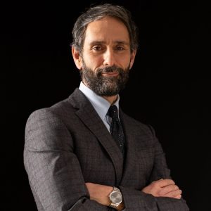
Antonio D’Amore, PhD (pictured), Research Assistant Professor in the Departments of Surgery and Bioengineering at the University of Pittsburgh, is leading a study funded through the European Research Council. The EU-funded BIOMITRAL project, which began in 2021, focuses on “tissue engineered valves that have the capacity to also regenerate the endogenous tissue, facilitate remodelling, and grow with the patient.” The project is hosted by the Fondazione RiMed in Italy and is being led by McGowan Institute affiliated faculty member, Antonio D’Amore, PhD, Research Assistant Professor in the Departments of Surgery and Bioengineering at the University of Pittsburgh.
McGowan Institute Director William Wagner, PhD, Distinguished Professor of Surgery, Chemical Engineering, and Bioengineering at the University of Pittsburgh, has been granted a subaward titled Human Heart Characterization and Nitinol Stent Fabrication. This subaward totals $97,073 and will last for one year.
The abstract of the BIOMITRAL grant reads:
Tissue Engineered Heart Valves (TEHVs) can restore function in the pulmonary and aortic positions and have shown capacity for tissue regeneration and growth in pre-clinical models. Yet, this concept has not been extended to the Mitral Valve (MV), whose pathologies affect >25% of the valve disease patients in Europe. In this proposal, we introduce a bio-inspired design methodology and bioprocessing technology to engineer BIOMITRAL: a polymeric, stent-less, tissue engineered MV that recapitulates native structure-function. Key to our approach is the engineering of MV leaflets and chordal apparatus. In the native MV, this set of tendon-like appendages mechanically connects the leaflets to the left ventricle (LV) and allows for harmonization of the valve kinematics, coaptation and ventricle contractile dynamics. Commercial MV prostheses used for MV replacement, as well as most existing TEHVs are mounted on synthetic stents that lack of this important structure and consequently neglect this physiological mechanism. In addition, non-degradable stents cannot adapt to patient’s growth, de-facto negating a key advantage in TEHVs. Our specific hypothesis is that recapitulating native leaflet structure-function and incorporating engineered chordal apparatus will lead to an engineered MV with enhanced functional and remodeling performances. To verify our hypothesis, we will: Aim 1. Characterize the structure-function of freshly isolated human valve tissue and use the derived properties to fabricate stented (control) and stentless BIOMITRAL prototypes; Aim 2. Assess prototypes mechanics and kinematics in silico via finite element modeling and in vitro in a pulse duplicator; Aim 3. Evaluate BIOMITRAL in vivo functional performance and assess remodeling in a chronic ovine model. Engineering a “living” MV with bioinspired leaflets and chordae that connect engineered leaflets with the LV is a revolutionary concept that can fundamentally transform the design of MV prostheses.
Congratulations Drs. D’Amore and Wagner!
RESOURCES AT THE MCGOWAN INSTITUTE
March Histology Special – Pentachrome Stain
Sprint into action and take advantage of our spring special at the McGowan Institute Histology Lab. As the days grow longer and warmer, nature begins to explode with color. The histology lab enjoys a little color too!
A Movat Pentachrome stain highlights the various components of connective tissue with five colors on a single stained slide.

Black=nuclei, Purple-black=elastic fibers, Red=Fibrinoid and muscle, Blue-blue green=ground or background substance, Yellow=reticulum fibers
You’ll receive 25% off Movat’s Pentachrome in March when you mention this ad.
Contact Julia at the McGowan Core Histology Lab by email: Hartj5@upmc.edu or call 412-624-5265.
Sample Submission Procedures: In response to COVID-19, we ask that you contact us to schedule a drop off time. When you arrive at the building you can call our laboratory at (412) 624-5365. Someone will meet you in the lobby to collect your samples. When your samples are completed, you will receive an email to schedule a pickup time.
UPCOMING EVENTS
Save the Date! 2023 McGowan Institute Scientific Retreat
![]()
The venue for the 2023 Retreat is the University Club, and the dates are March 6 & 7, 2023.
The program committee for the 2023 McGowan Institute Scientific Retreat, under the leadership of Bryan Brown, PhD, has begun to formulate the program and is requesting your suggestions for session topics.
To see the agenda and to register, please click here.
SCIENTIFIC ADVANCES
Bone Super Glue Returns to Space
RevBio, Inc., a Lowell, Mass.-based medical device company that makes a patented bone adhesive called Tetranite, which fixes fractures and accelerates bone repair—especially in patients suffering from osteoporosis, announced that an experiment to study this regenerative bone adhesive biomaterial has begun onboard the International Space Station (ISS). The study materials were launched on Saturday, November 26, 2022. This is RevBio’s second ISS mission.
RevBio’s first ISS mission was an in vitro experiment focused on the response of osteoblasts (a bone subtype) to Tetranite. This second mission is an in vivo experiment using Tetranite to treat 40 mice with artificially created bone defects while concurrently experimenting on 40 control mice on Earth to study the differences in microgravity. The experiment will examine the biomaterial’s osteoconductivity when used in a microgravity environment where bone density and the ability to regenerate new bone tissue is significantly compromised.
“This experiment is one of a kind, and will showcase the full potential of this breakthrough technology. The results will both validate and motivate our continued research efforts to commercialize products across several indications aimed at revolutionizing treatment options for patients with osteoporosis who suffer from debilitating fractures and have a poor prognosis for recovery,” said Brian Hess, co-inventor of the material and CEO of RevBio.
Principal Investigator and McGowan Institute affiliated faculty member, Giuseppe Intini, DDS, PhD (pictured), Associate Professor of Periodontics and Preventive Dentistry at the University of Pittsburgh, explains that the general theory is that microgravity impairs bone tissue regeneration by inhibiting differentiation of skeletal stem cells, and that Tetranite can reverse this inhibition and promote bone regeneration.
RevBio, Inc. was awarded a grant for this initial project through the Technology in Space Prize, funded by Boeing and the Center for the Advancement of Science in Space, Inc. (CASIS). The company is initially developing this technology for use in the dental market and recently initiated two clinical studies for the use of Tetranite to stabilize dental implants placed in sites that lack sufficient primary stability. The company is also working to develop adhesive applications for the broader orthopedics market.
Improving Vascular Grafts to Fight Cardiovascular Disease
Atherosclerosis – the buildup of fats, cholesterol and other substances in and on the artery walls, also known as plaque – can cause stroke, myocardial infarction and peripheral vascular disease. This increases the chances that a patient may die from cardiovascular disease.
Atherosclerosis can be treated through a vascular bypass procedure, where a synthetic graft is attached to a blood vessel and redirects blood flow around a blocked artery. In cases where patients have smaller or unusable blood vessels, a synthetic graft can fail due to blood clots or cells accumulating in the area.
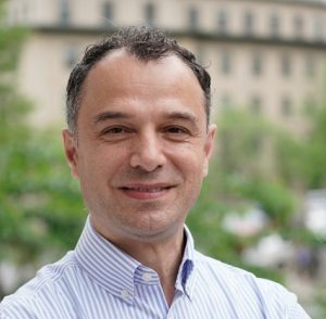
“These patients are in need of a bypass surgery,” said McGowan Institute affiliated faculty member Jonathan Vande Geest, PhD (pictured), professor of bioengineering at the University of Swanson School of Engineering. “The challenge is developing a synthetic graft that remains patent in both the short and long term.”
Dr. Vande Geest is part of a team of multidisciplinary researchers who are developing self-cleaning vascular grafts to support patients needing bypass surgery. These grafts would be able to repel microscopic blood cells called platelets, reducing the risk of a blood clot by preventing the formation of protein layers near the affected area.
Dr. Vande Geest and his team used a porohyperelastic model to increase the speed of the blood traveling radially inward from the luminal vessel wall to promote self-cleaning.
“This is just a start toward understanding self-cleaning grafts,” Dr. Vande Geest said. “More research will be needed to demonstrate improved functionality both on the bench and in appropriate preclinical animal models.”
The results of this research were published in the Journal of Biomechanical Engineering in February 2023.
Improving Motor Recovery in People with Chronic Post-Stroke Hemiparesis

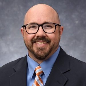
McGowan Institute for Regenerative Medicine affiliated faculty member Douglas Weber, PhD (pictured), Professor of Mechanical Engineering and Neuroscience at Carnegie Mellon University, is the co-principal investigator on the recently funded project entitled, “Spinal Cord Stimulation to Improve Motor Function in People with Post-Stroke Hemiplegia.” The 3-year project began on September 8, 2022, and was funded by the National Institute of Neurological Disorders and Stroke.
In the US almost 800,000 people have a stroke every year leaving more than half of these people with permanent arm and hand motor deficits (hemiparesis). Unfortunately, the only option available today is intense physical therapy, which improves motor performances but is not effective for many with moderate and severe post-stroke arm hemiparesis. In this project Dr. Weber and his team will test the efficacy of a novel neurostimulation system targeting the cervical spinal cord in combination with physical therapy to increase strength, ameliorate motor function, and improve motor recovery in people with chronic post-stroke hemiparesis.
The abstract of this project reads:
Motivation: In the US, almost 800,000 people have a stroke every year. Unfortunately, despite intense physio-therapy stroke survivors retain permanent arm motor deficits, some complete hemiparesis. Project Goal: Here we aim at testing the efficacy of a system delivering electrical neurostimulation to the cervical spinal cord (SCS) to improve arm and hand motor control in people with chronic post-stroke hemiparesis. Hypothesis: SCS of the lumbosacral circuits below the lesion immediately restored leg movements and weight bearing locomotion in humans with spinal cord injury by increasing strength and function. Moreover, SCS combined to physical therapy led to long-term improvements that improved motor control even when the stimulation was turned off. In stroke the spinal circuits controlling arm and hand movements are intact and located below the brain lesion. Therefore, we hypothesize that SCS targeting the cervical spinal cord will enable people with post-stroke hemiparesis to produce functional arm and hand movements, and that these improvements will enable the administration of a tailored behavioral training leading to superior long-term motor recovery. Preliminary data: We implanted a human subject with post-stroke hemiparesis with lateral cervical spinal leads and showed that SCS substantially improved, strength at every joint of the arm and hand, range of motions, dexterity, clinical assessment scales (Fugl-Meyer +13) and functional abilities. Moreover, SCS did not induce pain or discomfort at intensities that were necessary to obtain motor improvements. Finally, our industrial partner (CorTec GmbH), is finalizing the tests to obtain IDE approval of a fully implantable pulse generator tailored to SCS of the cervical spinal cord. Approach: In the UG3 phase of our proposal (years 1 to 3) we will finalize the necessary validation on our implantable cervical stimulator to obtain IDE approval. In parallel we will perform a pilot 30- day trial with conventional clinical leads used off-label in subjects with stroke to optimize SCS implant procedures and SCS parameters for maximization of arm functional movements. Finally, in the last 2 years during the UH3 phase we will execute an early feasibility study testing the efficacy of cervical SCS in combination with 4 weeks of physical therapy to improve arm motor control in up to 20 subjects with chronic post-stroke hemiparesis. Perspective: Our clinical trial will be the first to assess the efficacy of neurostimulation therapies targeting the spinal cord in patients with severe hemiparesis, potentially leading to a paradigm-shift in stroke rehabilitation.
Congratulations, Dr. Weber!
Review of Microfluidic Tools Shows Flow of Innovation

McGowan Institute for Regenerative Medicine affiliated faculty member Philip LeDuc, PhD (pictured), is a co-author of a new paper in Nature Communications entitled, “Microfluidics for Understanding Model Organisms.” The aims of this work are to encourage both engineers and biologists to develop microfluidics methods that can be widely used.
“I think oftentimes both fields can become siloed,” said Nolan Frey, a PhD candidate in Carnegie Mellon University’s (CMU’s) Department of Biological Sciences, a member of the Minden lab, and lead author on the review article. “Engineers tend to focus on tool design, whereas biologists focus on the biological systems they are investigating. But in my experience, it’s really the transdisciplinary collaboration between the two that allows for breakthroughs.”
Microfluidics involve transporting liquids through tiny channels that modulate fluid pressure to perform different tasks. They are used in a multitude of processes, from transferring ink when using a pen or watching liquid creep from a sample well in a COVID-19 test.
“Microfluidics, being the science and technology of systems that process or manipulate small amounts of fluids using microscopic channels, enables us to understand and manipulate different aspects of life at the very cellular level,” said Utku Sönmez, PhD, a recently graduated PhD candidate in mechanical engineering and a former member of CMU’s LeDuc lab. “This is a very recent and quite powerful ability for biological research.”
In biology, microfluidics is used to manipulate, immobilize, sort, and stimulate multicellular organisms. These methods can significantly decrease the amount of human handling necessary in experiments with living organisms, which is less stressful for the organisms and more accessible for human researchers. These methods also can be used on hundreds of organisms at once, saving time and allowing for a large sample size.
Mr. Frey uses microfluidics to study Drosophila (fruit fly) embryos, including how mutations affect the organisms’ gene expression and how they adapt to their environments. Microfluidics allow for precise pressure to be applied to the fly embryos, so Mr. Frey and fellow researchers can investigate how the embryos sense and respond to physical force.
“Microfluidics is allowing us to look at hundreds of embryos at a time,” said Jonathan Minden, PhD, professor of biological sciences at CMU. “We can do all sorts of different treatments to the embryos. Now, for the first time, we can learn what general gene expression changes there are.
Mr. Frey and Dr. Minden collaborate with researchers from CMU’s College of Engineering, including Drs. Sönmez and LeDuc, the William J. Brown professor of mechanical engineering, CMU, to design the tools necessary for his experiments.
“Using engineering techniques such as microfluidic systems to tackle biological problems inherently requires an interdisciplinary effort,” Dr. Sönmez said. “It may not be obvious how an engineering technique can enable new discoveries in biological sciences. However, if engineers and biologists communicate more frequently and chat about their interests and findings, there is a good chance that this will lead to unexpected developments.”
While pouring over articles on microfluidics and fruit flies, Mr. Frey and Dr. Sönmez compiled all the information they learned into a review article. They pitched the article to Nature Communications specifically as a review of how microfluidics can be used in fruit fly experiments. The editors thought the idea was too niche, and they suggested expanding the idea to include other species.
During the beginning and the height of the pandemic, while laboratory experiments were on hold, Mr. Frey and Dr. Sönmez read papers and wrote the review. Drs. LeDuc and Minden mentored them both during the process and served as additional authors.
Mr. Frey said one of the most promising aspects of microfluidics is that the necessary tools can be 3D printed with high-resolution equipment. This makes microfluidics less costly than other methods and means they can be created to suit more automated experiments.
“These technologies have gotten to the point where now you’re combining them with other systems, other new technologies like robotics,” Mr. Frey said. “It won’t be in the too distant future where we see basic experiments with model organisms — zebrafish, flies, etc. — also having the potential to be automated, and I think microfluidics is going to play a really key role in doing that.”
Mr. Frey said he hopes that microfluidics can be used in the new CMU Cloud Lab. These techniques would allow the first academic cloud lab to have a more diverse array of experiments.
“A big part of the reason I came to Carnegie Mellon was that I’m really interested generally in how technologies disrupt different fields, be it in research or more broadly in different aspects of society,” Mr. Frey said. “For me it’s been really cool and fun to participate in writing this article and to be part of this research with this team using new technologies on the cutting edge.”
Dr. LeDuc emphasized that this type of research is possible because of CMU’s emphasis on interdisciplinary partnerships.
“This type of project is the exact type of work that happens at Carnegie Mellon bringing together researchers from Biological Sciences and Engineering,” Dr. LeDuc said. “At some universities, they create centers and institutes to bring these types of researchers together, but at Carnegie Mellon it is in our blood and the way we think about everything.”
Carnegie Mellon Exosome Engineering Tech Licensed to Coya Therapeutics
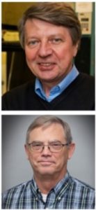
Carnegie Mellon University (CMU) has licensed a proprietary platform for bioengineering exosomes for drug delivery to Coya Therapeutics, Inc. Through the agreement, Coya has optioned the exclusive worldwide rights to research, develop, manufacture, and commercialize exosome polymer hybrids using the platform developed by an interdisciplinary team of CMU chemists and engineers. The team includes McGowan Institute for Regenerative Medicine affiliated faculty members Krzysztof Matyjaszewski, PhD (pictured top), professor of natural sciences, and Phil Campbell, PhD (pictured bottom), research professor of biomedical engineering.
Exosomes are a nanometer-sized type of extracellular vesicle that can be shed from every cell in the body and can hold a variety of cargo, including proteins, lipids, metabolites, and nucleic acids. Researchers worldwide have been attempting to harness the exosome as a drug delivery vehicle, but many of the techniques that have previously been developed required the use of complex molecular biology tools or degraded the exosome’s innate functionality.
Sushil Lathwal, PhD, then a doctoral candidate in the Mellon College of Science’s Department of Chemistry, and Saigopalakrishna (Sai) Yerneni, PhD, then a doctoral candidate in the College of Engineering’s Department of Biomedical Engineering, came up with an idea to surmount these obstacles. They worked alongside researchers in the laboratories of Subha Das, PhD, associate professor of chemistry, Dr. Matyjaszewski, and Dr. Campbell, to create a method that engineers exosomes with a DNA-cholesterol tether. The synthetic single-stranded DNA on the tether can bind with a complementary strand of DNA linked to a bioactive agent.
“Our research on exosome surface modification started, in the best traditions of Carnegie Mellon University, as an interdisciplinary collaborative project. Although this project began out of simple curiosity of combining our respective research interests, we are delighted to see its potential clinical translation with our venture with Coya Therapeutics,” Drs. Das and Campbell said in a statement.
Coya Therapeutics is a clinical-stage biotechnology company that develops approaches that enhance regulatory T cell (Tregs) in vivo, including autologous and allogenic Treg-derived exosome therapeutic and novel biologics. They plan to develop exosome-polymer hybrids (EPHs) to enable exosomes to be homed to proteins of interest, while delivering select payloads to targeted cells.
AWARDS AND RECOGNITION
Dr. Tracy Cui Named to 2023 Class of NAI Senior Members
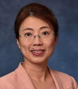
The National Academy of Inventors (NAI) has named 95 of the world’s best emerging academic inventors to its 2023 class of Senior Members. One of those honorees is McGowan Institute for Regenerative Medicine faculty member Xinyan Tracy Cui, PhD (pictured), William Kepler Whiteford Professor of Bioengineering at the University of Pittsburgh.
NAI Senior Members are active faculty, scientists, and administrators from NAI Member Institutions who have demonstrated remarkable innovation producing technologies that have brought, or aspire to bring, real impact on the welfare of society. They also have growing success in patents, licensing, and commercialization.
“Tracy Cui’s induction into the National Academy of Inventors is a well-deserved recognition of her innovative spirit and exceptional contributions to the field of biomedical engineering,” said Mark Redfern, interim chair of the Department of Bioengineering and William Kepler Whiteford Professor. “Her dedication to creating technologies that improve human health is an inspiration to all aspiring inventors, and we look forward to the many breakthroughs she will undoubtedly achieve in the years to come.”
The NAI’s mission is to recognize and encourage inventors with patents issued from the U.S. Patent and Trademark Office, enhance the visibility of academic technology and innovation, encourage the disclosure of intellectual property, educate, and mentor innovative students, and translate the inventions of its members to benefit society. The Senior Member Program allows the NAI to expand its reach and further its goals in recognizing the contributions of inventors around the globe.
Congratulations, Dr. Cui!
Dr. Rory Cooper Inducted into the National Inventors Hall of Fame
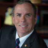
McGowan Institute affiliated faculty member Rory Cooper, PhD (pictured), Distinguished Professor of the Department of Rehabilitation Science & Technology, and professor of Bioengineering, Physical Med & Rehab, and Orthopedic Surgery at the University of Pittsburgh, as well as Founding Director and VA Senior Research Career Scientist of the Human Engineering Research Laboratories (HERL), has been named to the 50th class of the National Inventors Hall of Fame for his inventions in mobility technology to aid wheelchair users.
Dr. Cooper founded HERL to solve common problems faced by disabled veterans and other wheelchair users. Herl is a part of the U.S. Department of Veterans Affairs as the Center for Wheelchairs and Assistive Robotics Engineering. Herl aims to create a world where all people with disabilities have unencumbered mobility and function so that they can fully participate in and contribute to society.
Recently, Dr. Cooper was featured in Next Pittsburgh.
The National Inventors Hall of Fame, now celebrating its 50th year, aims to advance the spirit of innovation by inspiring emerging creators and entrepreneurs through their education programs and honoring the history of innovation through their museum and Hall of Fame Inductees.
Dr. Cooper will be honored along with other inductees at a celebration in Washington, D.C., on Oct. 26, 2023.
Congratulations, Dr. Cooper!
Two Bioengineering Graduate Students Receive American Heart Association Fellowships
Two students from the labs of McGowan affiliated faculty members in the University of Pittsburgh Department of Bioengineering received American Heart Association Predoctoral Fellowships, which provides up to three years of project support for aspiring academic and health professionals related to cardiovascular research.
“We are delighted to see our students receive the coveted American Heart Association predoctoral fellowships. This is a testament to their hard work, dedication, and passion for creating knowledge in the cardiovascular arena and advancing the fight against heart disease and stroke,” said McGowan Institute faculty member Sanjeev Shroff, PhD, Distinguished Professor and Gerald E. McGinnis Chair of Bioengineering and Interim U.S. Steel Dean of Engineering. “This is the largest number of AHA fellowships the Department of Bioengineering PhD students have received in a single year, and I am proud of the research accomplishments of each of these students. I look forward to seeing the continued growth of these students as independent investigators.”
The AHA predoctoral fellows are:
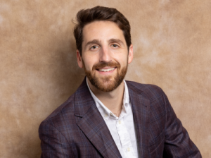 Matthew Borrelli (PIs: McGowan Institute faculty member Steven R. Little, PhD and Hēth Turnquist, PhD)
Project: Local Enhancement of Regulatory T Cells for the Treatment of Myocardial Infarction
Mr. Borrelli’s project focuses on developing a new way to address the damage caused by the immune system after a heart attack. Regulatory T cells (Treg) are used to control inflammation in the body. Previous research has discovered a process to increase the number of Treg by delivering proteins that can attract or induce Treg, which reduces inflammation. Mr. Borrelli will attempt to expand on this by shifting the focus to an injectable technology.
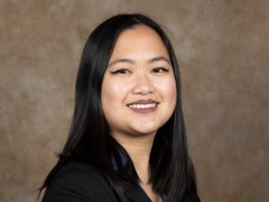 Andi Marini (PI: McGowan Institute affiliated faculty member Justin Weinbaum, PhD)
Project: Extracellular vesicle delivery system for treatment for Abdominal Aortic Aneurysm
Ms. Marini’s project focuses on the development and characterization of a localized controlled release system for adipose stem cell derived extracellular vesicles (EVs). The project experimentally and computational analyzes EV diffusion into aortic tissue. The goal is fabrication of a local periadventitial abdominal aortic aneurysm (AAA) treatment. If successful, the results will lay the groundwork for a novel regenerative treatment for small AAAs to prevent pre-threshold rupture and halt aneurysm growth.
Congratulations, Mr. Borrelli and Ms. Marini!
Illustration: University of Pittsburgh Swanson School of Engineering.
PUBLICATION OF THE MONTH
Author: Ali Behrangzade, Bruce R. Simon, William R. Wagner, Jonathan P. Vande Geest
Title: Optimizing the Porohyperelastic Response of a Layered Compliance Matched Vascular Graft to Promote Luminal Self-Cleaning
Summary: Thrombosis and intimal hyperplasia have remained the major failure mechanisms of small-diameter vascular grafts used in bypass procedures. While most efforts to reduce thrombogenicity have used a biochemical surface modification approach, the use of local mechanical phenomena to aid in this goal has received somewhat less attention. In this work, the mechanical, fluid transport, and geometrical properties of a layered and porous vascular graft are optimized within a porohyperelastic finite element framework to maximize self-cleaning via luminal reversal fluid velocity (into the lumen). This is expected to repel platelets as well as inhibit the formation of and/or destabilize adsorbed protein layers thereby reducing thrombogenic potential. A particle swarm optimization algorithm was utilized to maximize luminal reversal fluid velocity while also compliance matching our graft to a target artery (rat aorta). The maximum achievable luminal reversal fluid velocity was approximately 246 μm/s without simultaneously optimizing for host compliance. Simultaneous optimization of reversal flow and compliance resulted in a luminal reversal fluid velocity of 59 μm/s. Results indicate that a thick highly permeable compressible inner layer and a thin low permeability incompressible outer layer promote intraluminal reversal fluid velocity. Future research is needed to determine the feasibility of fabricating such a layered and optimized graft and verify its ability to improve hemocompatibility.
Source: J Biomech Eng. Feb 2023, 145(2): 021002.
GRANT OF THE MONTH
PI: William Wagner
Title: Human Heart Characterization and Nitinol Stent Fabrication
Description: Tissue Engineered Heart Valves (TEHVs) can restore function in the pulmonary and aortic positions and have shown capacity for tissue regeneration and growth in pre-clinical models. Yet, this concept has not been extended to the Mitral Valve (MV), whose pathologies affect >25% of the valve disease patients in Europe. In this proposal, we introduce a bio-inspired design methodology and bioprocessing technology to engineer BIOMITRAL: a polymeric, stent-less, tissue engineered MV that recapitulates native structure-function. Key to our approach is the engineering of MV leaflets and chordal apparatus. In the native MV, this set of tendon-like appendages mechanically connects the leaflets to the left ventricle (LV) and allows for harmonization of the valve kinematics, coaptation and ventricle contractile dynamics. Commercial MV prostheses used for MV replacement, as well as most existing TEHVs are mounted on synthetic stents that lack of this important structure and consequently neglect this physiological mechanism. In addition, non-degradable stents cannot adapt to patient’s growth, de-facto negating a key advantage in TEHVs. Our specific hypothesis is that recapitulating native leaflet structure-function and incorporating engineered chordal apparatus will lead to an engineered MV with enhanced functional and remodeling performances. To verify our hypothesis, we will: Aim 1. Characterize the structure-function of freshly isolated human valve tissue and use the derived properties to fabricate stented (control) and stentless BIOMITRAL prototypes; Aim 2. Assess prototypes mechanics and kinematics in silico via finite element modeling and in vitro in a pulse duplicator; Aim 3. Evaluate BIOMITRAL in vivo functional performance and assess remodeling in a chronic ovine model. Engineering a “living” MV with bioinspired leaflets and chordae that connect engineered leaflets with the LV is a revolutionary concept that can fundamentally transform the design of MV prostheses.
Source: RiMed
Term: One year
Amount: $97,073