February 2022 | VOL. 21, NO. 2| www.McGowan.pitt.edu
New Peer-Reviewed Publication Expanding Liver Regeneration Technology

McGowan Institute for Regenerative Medicine faculty member Eric Lagasse, PharmD, PhD, is an Associate Professor in the Department of Pathology at the University of Pittsburgh and a co-founder and Chief Scientific Officer of LyGenesis, a clinical-stage biotechnology company developing cell therapies for patients with end stage liver disease, Type 1 diabetes, end stage renal disease, single enzyme deficiencies, and aging.
A decade ago, Dr. Lagasse, a world leader in ectopic transplantation research conducted at his McGowan Institute laboratory, began a series of experiments that would form the foundation for LyGenesis. He discovered that hepatocytes (liver cells) transplanted into lymph nodes would not just survive, but thrive, organize, and begin to function as miniature ectopic livers, exerting life-saving effects in otherwise fatal small and large animal models of end stage liver disease. His research confirms that it is possible to harness the body’s lymph nodes as bioreactors for growing functional ectopic organs.
Dr. Lagasse and McGowan Institute affiliated faculty member Maria Giovanna Francipane, PhD, Principal Investigator in Regenerative Medicine at Fondazione Ri.MED (Palermo, Italy), an international partnership between the Italian Government, the Region of Sicily, the Italian National Research Council (CNR), the University of Pittsburgh, and UPMC, are co-authors on the publication of a new peer-reviewed paper published in the journal Hepatology on LyGenesis’s organ regeneration technology.
LyGenesis’s technology is built on a broad foundation of small and large animal preclinical studies showing that lymph nodes can serve as sites for organogenesis – the growth of functioning ectopic organs. This latest publication entitled “Fat-Associated Lymphoid Clusters as Expandable Niches for Ectopic Liver Development,” demonstrates that another lymphoid site, the fat-associated lymphoid cluster, can act as an additional anatomical location for ectopic liver development and restoration of liver function.
“This study consolidates and expands on the use of lymphoid sites, including the lymph node and fat-associated lymphoid clusters, for the development of functional ectopic livers. In this latest study, we were able to demonstrate the compensatory role of ectopic livers grown within the fat-associated lymphoid clusters, which resulted in the restoration of liver function and rescuing the animals from otherwise fatal liver disease,” said Dr. Lagasse.
David Roblin, MBBS, FMedSci, CEO of Juvenescence Therapeutics, a lead investor in LyGenesis, noted, “This latest peer-reviewed publication in one of the top journals in the field further supports LyGenesis’s platform technology using the lymph node and now other lymphoid sites as bioreactors to grow functional, life-saving tissues. We are excited about the potential to impact patients as LyGenesis commences its first clinical trial among patients with end stage liver disease.”
LyGenesis’s lead allogeneic cell therapy program is currently in a Phase 2a clinical trial for patients with end stage liver disease (ClinicalTrials.gov Identifier: NCT04496479).
RESOURCES AT THE MCGOWAN INSTITUTE
March Histology Special – Pentachrome Stain
Sprint into action and take advantage of our spring special at the McGowan Institute Histology Lab. As the days grow longer and warmer, nature begins to explode with color. The histology lab enjoys a little color too!
A Movat Pentachrome stain highlights the various components of connective tissue with five colors on a single stained slide.

Black=nuclei; Purple-black=elastic fibers; Red=Fibrinoid and muscle; Blue-blue green=ground or background substance; Yellow=reticulum fibers
You’ll receive 25% off Movat’s Pentachrome stains in March when you mention this ad.
Contact Julia at the McGowan Core Histology Lab by email: Hartj5@upmc.edu or call 412-624-5265.
New Sample Submission Procedures: In response to COVID-19, we ask that you contact us to schedule a drop off time. When you arrive at the building you can call our laboratory at (412) 624-5365. Someone will meet you in the lobby to collect your samples. When your samples are completed, you will receive an email to schedule a pickup time.
Save the Date-2022 McGowan Institute Scientific Retreat
 Now Open for the 2022 McGowan Institute Scientific Retreat
The 21st Annual McGowan Institute for Regenerative Medicine Virtual Scientific Retreat will take place on March 7-8, 2022.
The poster session will be held on March 7th.
Under the leadership of McGowan Institute for Regenerative Medicine faculty member Bryan Brown, PhD, associate professor, Department of Bioengineering the program committee is planning an exciting group of speakers and topics.
Mark the dates on your calendars. Additional 2022 Retreat details and news are available here.
SCIENTIFIC ADVANCES
Calming the Cytokine Storm
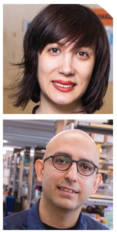
When her father was diagnosed with pancreatic cancer, McGowan Institute for Regenerative Medicine affiliated faculty member Samira Kiani, PhD, Associate Professor, Liver Research Center, Department of Pathology at the University of Pittsburgh, scoured available clinical trials for cutting edge gene and cell therapies but came away frustrated at the limitations posed by many of them as a result of their vulnerability to the body’s natural immune response to the therapies.
That frustration has sharpened her focus as a biomedical researcher, beginning as a postdoctoral fellow at the Massachusetts Institute of Technology, where she began exploring ways to make the CRISPR-Cas9 gene editing technology more safe and more effective.
Dr. Kiani moved on to Arizona State University to start her own lab where she honed-in on the concept of using a modified version of CRISPR to dial up and down expression of genes that regulate immune response to therapy.
“If you put a pathogen into a human the immune system will react. My focus was on how to make a safer CRISPR-based gene therapy,” she said. “We picked a gene involved in immune response to explore how to modulate the reaction to CRISPR, as well as using CRISPR to modulate the immune response.”
Something was missing in her lab, however, that she felt was hindering the potential of her research – access to clinicians and patients. That’s when she and her primary collaborator, McGowan Institute affiliated faculty member Mo Ebrahimkhani, MD, Associate Professor, Liver Research Center, accepted an offer to create a lab at the University of Pittsburgh in the Department of Pathology within the School of Medicine, which enjoys a clinical collaboration with the UPMC health system.
She was also attracted by what she saw as Pitt’s emerging culture of innovation and entrepreneurship, as she had developed an interest in commercializing her lab’s discoveries so that they can have an impact on the treatment of disease.
Six months ago, she formed GenExGen, a company that intends to bring to market a new generation of immune suppressive therapies using CRISPR, with herself as the CEO. The company remains virtual for now as Dr. Kiani works to build its foundation. She put together a pitch deck to begin approaching potential investors.
“It has been a steep learning curve. They (investors) give you all these questions that you don’t think about from a research perspective. I immediately got a lot of feedback that investors did not want to fund an ‘add-on’ technology that supplemented or enabled other therapies. They wanted a stand-alone therapeutic,” she said.
Based on pre-clinical data in mice, Dr. Kiani said her lab discovered one immediate indication to suppress immune response to treat a dangerous condition called “ the cytokine storm” in which the body’s immune response to a pathogen or medical treatment goes into overdrive producing a severe inflammatory response, potentially causing organ damage or even death. They are also examining the impact of this approach in modulating immune response to the gene therapies of cystic fibrosis.
“Later in 2022, once we have more data, we will look to raise an investment round, or partner with other companies to fund studies that lead to an investigative new drug filing with the FDA to begin the clinical trial process,” she said, while also exploring additional federal research and commercialization funding, such as the Small Business Innovation Research (SBIR) or Small Business Technology Transfer (STTR) programs.
Eventually Dr. Kiani said she would be interested in hiring an “industry savvy” CEO to navigate the journey to market, allowing her to potentially transition into a chief science officer role, while she continues her research into building new tools for cell and gene therapies.
“Having translational experience is making me a better researcher,” she said. “It informs how you run your research program in your lab and helps you be able to write better grant applications.”
Characterizing the Progression of Atherosclerotic Plaques and Predicting Rupture Vulnerability

Acute coronary syndromes and strokes together constitute a leading cause of morbidity and mortality in the United States and Europe, approximately 80% of which are caused by atherosclerotic plaque rupture. Validation of an innovative diagnostic ultrasound imaging technology that will allow for noninvasively detecting atherosclerotic plaques that have high chance of rupturing, which will result in heart attacks and strokes, is proposed.
McGowan Institute for Regenerative Medicine affiliated faculty member Kang Kim, PhD, Associate Professor of Medicine and of Bioengineering at the University of Pittsburgh and the Heart and Vascular Institute, UPMC, is the principal investigator on the project entitled, “Super Resolution Ultrasound Imaging of Vasa Vasorum to Characterize the Progression of Atherosclerotic Plaques and Predict Rupture Vulnerability,” which will run for 4 years. The NIH National Heart, Lung, and Blood Institute funded this work.
The innovative technology, so called super resolution ultrasound imaging, provides unprecedented high resolution that enables imaging abnormal growth of microvessels in atherosclerotic plaques at great details, which is well known as a key indicator of plaque rupture. If successfully validated through a novel rabbit plaque rupture model that is known to be most close to human plaques, the technology development and findings through this project will provide strong foundation and potential for future clinical translation. Upon successful clinical validation in the future on human subjects, an immediate clinical application will be for carotid atherosclerosis clinics where this imaging technology incorporated into a current ultrasound scanner will be able to identify plaques at high risk of rupture for most appropriate interventions and/or treatments to prevent strokes and save life. The immediate outcomes from this project include an accurate, affordable, well-validated robust animal imaging tool for important diseases which are associated with microvessel abnormalities such as atherosclerotic plaques proposed in this project, cancer angiogenesis, and kidney diseases to name a few.
The abstract for this project follows:
Acute coronary syndromes and strokes together constitute a leading cause of morbidity and mortality in the United States and Europe, approximately 80% of which are caused by atherosclerotic plaque (AP) rupture. Over the past decade, extensive efforts have been made to identify a rupture-prone AP. Among others, infiltration of dense neovascularization arising from vasa vasorum (VV) into the AP core plays a critical role in AP rupture. Postmortem studies revealed key involvement of VV in AP. However, a persistent lack of a noninvasive, high-resolution imaging tool to longitudinally assess abnormal microvascular expansion remains a critical barrier to adequate in-vivo investigation on how VV affects AP progression and contributes to eventual rupture.
To address this dire unmet need, we propose an innovative transcutaneous super resolution ultrasound (SRU) imaging. The technology development in this project seeks to shift the current US imaging approach in identifying microvessels of AP from “intravascular” to a “fully noninvasive transcutaneous” imaging approach. This is only possible by achieving unprecedented high spatial resolution at large depth, breaking acoustic diffraction limit of the ultrasound frequency that governs spatial resolution. Our group has performed in-depth feasibility studies where SRU imaging successfully identified neomicrovessels in cholesterol-fed rabbit AP, evaluated against µCT and histology. Additionally, areas requiring further technical optimization were identified. Such technology developments and preliminary data thus far rigorously support our overarching hypothesis that enhanced and optimized SRU will accurately stage plaque progression and identify rupture-prone plaques by directly measuring VV changes with exquisite detail. To test the hypothesis, we will use a well-established, clinically relevant cholesterol-fed rabbit AP rupture model, which has shown the most similarity to human plaque pathology including VV neovascularization, to validate the novel SRU system to 1) Successfully quantify changes in vessel density and 2) Identify rupture-prone AP.
To achieve these goals, we propose the following specific aims: 1) To develop enhanced SRU at high frequency using a commercial small animal imaging probe, and 2) To determine if VV changes estimated by SRU correlate with AP progression and are predictive of AP rupture. The immediate outcomes of the proposed work are an affordable noninvasive small animal SRU imaging tool and it’s validation on a clinically relevant rabbit AP model, which also can be used for other important small animal disease models, which are associated with microvessel abnormality such as cancer angiogenesis and kidney diseases to name a few. With proper adaptations into a clinical mid frequency probe and validation in clinical settings in future, this work will lead to our long-term translational goal to integrate SRU in a facile manner into the current clinical standard of carotid duplex sonography that has shown poor specificity to plaque vulnerability. This will help to effectively stratify patients at high risk of strokes and guide adequate intervention/treatment options for stroke prevention, exerting highly influential clinical impact.
New Computational Tool Predicts Cell Fates and Genetic Perturbations
Imagine a ball thrown in the air: It curves up, then down, tracing an arc to a point on the ground some distance away. The path of the ball can be described with a set of simple mathematical equations, and if you know the equations, you can figure out where the ball is going to land.
Biological systems tend to be harder to forecast, but researchers from the University of Pittsburgh School of Medicine and collaborators at Whitehead Institute for Biomedical Research are working on making the path taken by cells as predictable as the arc of a ball. Rather than looking at how cells move through space, they are considering how the state of a cell, defined by the expression levels of its genes, evolves with time.
 In a new paper published in the journal Cell, Jianhua Xing, PhD, Professor of Computational and Systems Biology at Pitt, and collaborators describe a computational framework – named Dynamo — that allows them to predict a cell’s fate without performing laborious and costly experiments at the lab bench. Dr. Xing’s team includes McGowan Institute for Regenerative Medicine affiliated faculty member Ivet Bahar, PhD, Distinguished Professor, the John K. Vries Chair, and the Founding Chair in the Department of Computational and Systems Biology at the University of Pittsburgh School of Medicine, and student Yan Zhang.
“A cell is a dynamical system that receives and processes a lot of information from the surrounding environment and responds accordingly through a complex regulatory network of genes,” said Dr. Xing. “If we devise a set of equations that can describe how genes within a cell regulate each other, we can computationally predict how to transform terminally differentiated cells into stem cells or how a cancer cell may respond to various combinations of drugs that would be impractical to test experimentally.”
Along with their collaborators Jonathan Weissman, PhD, and Xiaojie Qiu, PhD, at Whitehead Institute, the Pitt team has developed a machine-learning framework that can construct the mathematical equations describing a cell’s trajectory from one state to another.
The framework also can be used to figure out the underlying mechanisms—the specific cocktail of gene activity—driving changes in the cell. And the researchers hope that Dynamo will not only help them understand how cells transition from state to state, but also guide them in controlling this process.
Dynamo includes tools to simulate how a cell’s gene expression behaviors change based on different manipulations, and a method to find the most efficient way to reprogram any cell type to another — a fundamental challenge in stem cell biology and regenerative medicine — as well as help generate hypotheses of how genetic changes will collectively alter a cell’s fate.
“Our goal is to move towards a more quantitative version of single cell biology,” said Dr. Qiu. “We want to be able to map how a cell changes in relation to the interplay of regulatory genes as accurately as an astronomer can chart a planet’s movement in relation to gravity, and then we want to understand and be able to control those changes.”
Youbiotics Receives Commercialization Grant from Pitt Chancellor’s Gap Fund
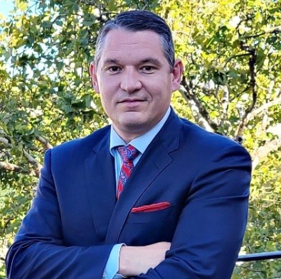
Four Pitt innovation teams—including one headed by McGowan Institute for Regenerative Medicine faculty member Steven Little, PhD, a Distinguished Professor, the Chairman of the Department of Chemical and Petroleum Engineering, and the William Kepler Whiteford Endowed Professor in the Departments of Chemical and Petroleum Engineering, Bioengineering, Immunology, Ophthalmology, and Pharmaceutical Sciences—have been selected to receive commercialization grants from the Chancellor’s Gap Fund in order to accelerate their path from the lab to the market.
Details of Dr. Little’s funded project follow:
Youbiotics
- Project Description: Personalized probiotics for weight management.
- Principal Investigator: Steven Little, PhD, Swanson School of Engineering.
- Co-Investigators: Matt Borrelli, Swanson School of Engineering; Abhinav Acharya, PhD, Arizona State University; Jonathan Krakoff, MD, National Institutes of Health.
The Chancellor’s Gap Fund was reauthorized in 2021 to provide critical bridge funding for research projects that have demonstrated strong commercialization potential but require key proof of concept experiments or other data or prototypes in order to attract interest from potential investors or industry partners.
The fund provides grants ranging from $25,000 to $75,000, based on what is needed to advance the project through a significant milestone. A total of $263,000 was awarded in this cycle.
“The standard of applications remains consistently high, and we look forward to helping these teams not only reach their program goals, but also move along the path to creating significant social and financial impact as a result,” said Peter Allen, PhD, Executive Director, Innovation and Strategy, Office of Innovation and Entrepreneurship.
Can AI Help Unravel the Mysteries of Sepsis and Save Lives?
Researchers are investigating the use of artificial intelligence to forecast therapeutic effectiveness and outcomes for patients with sepsis, a syndrome that claims one in five lives around the world and has until now been a black box for rapid diagnosis and treatment.
In the United States, more than a quarter of a million people each year succumb to sepsis, which occurs when the immune system responds to an existing infection such as COVID-19 by turning on itself instead of fighting the germs.
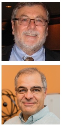
The research team, led by Rishikesan Kamaleswaran, PhD, assistant professor in Emory’s biomedical informatics department, was recently awarded a $2.6 million award from the National Institute of General Medical Sciences of the National Institutes of Health to mine sensor-generated data streams for physio-markers that may be able to predict the onset of sepsis, inform treatment options, and support the discovery of sub-types of sepsis. McGowan Institute for Regenerative Medicine affiliated faculty members Michael Pinsky, MD, Professor of Critical Care Medicine, Bioengineering, Cardiovascular Diseases, Clinical & Translational Science, and Anesthesiology at the University of Pittsburgh, and Gilles Clermont, MD, Professor of Critical Care Medicine, Industrial Engineering, and Mathematics at the University of Pittsburgh, are members of the project’s research team.
“The hope is to use AI in a new way so we can better see in an area where traditionally we have flown blind,” says Dr. Kamaleswaran. “By using existing and routinely collected physiological data in intensive care units (ICU), we hope to generate robust machine learning algorithms than can more 1. More accurately phenotype sepsis patients and 2. Improve outcomes with more personalized treatments – therapies that would elicit the best response.”
Approaches using machine learning have focused largely for predicting sepsis from electronic medical records which Dr. Kamaleswaran says suffer from a host of problems. “The data are not timely, significant portions are missing or wrong because of the manual process of entry, and the information often reflects individual and institutional biases, which all make it difficult to devise a treatment plan that can be replicated someplace else.”
The five-year study will tap into expertise from different disciplines at Emory including mathematics, computer science, and medicine to develop sophisticated tools that can analyze the data, identify patterns, and prescribe a course of action. “We will contribute significant knowledge about the role and utility of complex physiological interactions that are abundantly available in clinical practice but seldom used for clinical decision making.”
Dr. Kamaleswaran points out that sepsis results in many deaths from COVID-19 and other diseases. “The use of AI and machine learning here are powerful mathematical constructs that when placed in the hands of a capable clinician, can become an efficient resource for improving patient care.”
The tools the research team is developing rely on information from patients who developed secondary infections after admission to the ICU, a departure from existing algorithms for sepsis that use data from patients in general wards.
Apart from Drs. Kamaleswaran, Pinsky, and Gilles, the research team for the study comprises physician Craig Coopersmith, biomedical informatics scientists Gari Clifford and Qiao Li, all at Emory; and scientist Omer Inan at the Georgia Institute of Technology.
Infections that lead to sepsis most often start in the lung, urinary tract, skin, or gastrointestinal tract. Without timely treatment, sepsis can rapidly lead to tissue damage, multi-organ failure, and death.
The Centers of Disease Control and Prevention says one in three patients who die in a hospital in the U.S has sepsis, and in 87 percent of those cases, the infection begins outside the health care facility.
The World Health Organization has said that lowering the death and disability count from sepsis is particularly challenging because of “serious gaps in knowledge” particularly in low- and middle-income countries. Around 11 million people die each year of sepsis, many of them children, and even those who survive can sometimes have lifelong disabilities.
“The goal is to find a novel way to improve patient care and move the needle in a space that is plagued with complexity,” says Dr. Kamaleswaran.
Researchers Discover New Way to Target Secondary Breast Cancer that Has Spread to the Brain
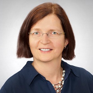
An international collaborative study by researchers at Royal College of Surgeons in Ireland (RCSI) University of Medicine and Health Sciences and the Beaumont RCSI Cancer Centre (BRCC), the Mayo Clinic, and the University of Pittsburgh has revealed a potential new way to treat secondary breast cancer that has spread to the brain, using existing drugs. The study is published in Nature Communications.
The research was led by BRCC investigators Professor Leonie Young, Dr. Nicola Cosgrove, Dr. Damir Varešlija, and Professor Arnold Hill. McGowan Institute for Regenerative Medicine affiliated faculty member Steffi Oesterreich, PhD, the Shear Family Foundation Chair in Breast Cancer Research Professor and Vice-Chair Department of Pharmacology and Chemical Biology, University of Pittsburgh; Co-Leader Cancer Biology Program, UPMC Hillman Cancer Center (UPMC HCC); and Co-Director Women’s Cancer Research Center, UPMC HCC and Magee Womens Research Institute, is a co-author of the study.
Most breast cancer related deaths are a result of treatment relapse leading to spread of tumors to many organs around the body. When secondary breast cancer, also known as metastatic breast cancer, spreads to the brain it can be particularly aggressive, sometimes giving patients just months to live.
The RCSI study focused on genetically tracking the tumor evolution from diagnosis of primary breast to the metastatic spread in the brain in cancer patients. The researchers found that almost half of the tumors had changes in the way they repair their DNA, making these tumors vulnerable to an existing type of drug known as a PARP inhibitor. PARP inhibitor drugs work by preventing cancer cells to repair their DNA, which results in the cancer cells dying.
“There are inadequate treatment options for people with breast cancer that has spread to the brain and research focused on expanding treatment options is urgently needed. Our study represents an important development in getting one step closer to a potential treatment for patients with this devastating complication of breast cancer,” commented Professor Leonie Young, the study’s Principal Investigator.
“By uncovering these new vulnerabilities in DNA pathways in brain metastasis, our research opens up the possibility of novel treatment strategies for patients who previously had limited targeted therapy options,” said study author Dr. Damir Varešlija.
When a Protective Gene Buffers a Bad One, a Heart Can Beat
It was a medical mystery: When University of Pittsburgh School of Medicine scientists induced a particular genetic mutation in mouse eggs, the resulting embryos would all die in the womb within a week.
And yet, people with the same troublesome gene are thriving.
“This gene is clearly very deleterious – the mice did not even develop a heartbeat, let alone survive to birth,” said Cecilia Lo, PhD, Distinguished Professor and F. Sargent Cheever Chair of Pitt’s Department of Developmental Biology. “That led us to wonder: How are people who we know have this gene walking around?”
 The team—including McGowan Institute for Regenerative Medicine affiliated faculty member Kyle Orwig, PhD, Professor, OB/GYN and Reproductive Sciences, University of Pittsburgh School of Medicine, and the founding Director of the Fertility Preservation Program of UPMC and the UPMC Magee Center for Reproduction and Transplantation—found that a protective gene was countering the bad one, explaining why some people with this very deleterious gene not only survived but did so with only an atrial septal defect—a hole in the heart. The findings—reported in Cell Reports Medicine—provide valuable clinical and personal information to guide families with a history of the disease and could lead to future genetic treatments.
Congenital heart disease is one of the most common birth defects, affecting about 1% of live births. Atrial septal defects—which involve a hole in the wall between the upper chambers of the heart, allowing blood to flow in ways that can damage the heart and lungs—are among the most common forms of congenital heart disease, affecting as many as 10,000 babies born in the U.S. each year.
Working with Brian Feingold, MD, MS, medical director of the pediatric heart failure and heart transplant programs at UPMC Children’s Hospital of Pittsburgh, Dr. Lo’s team obtained genetic samples from eight members of a family who all had large atrial septal defects. Whole genome sequencing revealed that they all carried an extremely rare mutation in a gene called TPM1 that didn’t appear in more than 900 unrelated samples from people with congenital heart disease; worldwide, it has been seen only twice.
To learn more about this genetic mutation, Dr. Lo’s team used CRISPR-Cas9 gene editing to introduce this mutation in mouse embryos. The embryos would develop normally to about 8.5 days—exactly when the heart should start to beat—and then die without a heartbeat. Co-lead author Xinxiu “Cindy” Xu, PhD, a postdoctoral associate at Pitt, determined that the TPM1 mutation was inhibiting production of a protein essential for heart beating.
The mouse deaths and the extreme rarity in humans indicated that nobody with this mutation should live, but since the scientists had genetic samples from eight related people with beating hearts, Dr. Xu created a “disease-in-a-dish” model to see what would happen with the patient cells in a petri dish. This confirmed the patient-derived heart cells beat normally.
So, Dr. Lo’s team knew there had to be more to the story. That’s when they started to suspect a protective gene was at play. They went back to the drawing board and further scoured the genomic sequences.
“With the modern tools we have for exploring genetics, we’re learning that not everything is as it seems,” Dr. Lo said. “There are complexities that are important for our understanding of the genetic etiology of disease.”
Focusing on the same three sections of the chromosome inherited by all eight family members, Dr. Lo’s team found nine additional genetic variations in close proximity to the bad TPM1 mutation. Only one—TLN2—was co-expressed with TPM1 in the cardiac cells responsible for making the heart beat.
When the team introduced both the deleterious and protective mutations simultaneously in the mouse embryos, beating hearts were observed. Atrial septal defect still occurred, as seen in the family members with these two mutations. The protective gene wasn’t strong enough to completely overpower the damage caused by the bad one, but the heart could beat well enough to sustain life.
The discovery can have immediate implications for helping families understand the risk of passing the mutations to future generations, as well as help guide clinical treatment, such as prompting doctors to consider early treatments or more frequent assessments for heart dilation and rhythm disturbances. For example, with some heartbeat irregularities, surgically implanting a pacemaker or a defibrillator could be helpful in restoring normal heart beating.
Scientific advances also can uncover big picture implications, Dr. Lo said. “The future of genetic therapy doesn’t have to be about turning off bad genes. It can also be about turning on good ones.”
Dr. Tagbo Niepa Receives 2022 NSF CAREER Award

The National Science Foundation (NSF) has awarded a CAREER Award to McGowan Institute for Regenerative Medicine affiliated faculty member Tagbo Niepa, PhD, Assistant Professor in the Department of Chemical and Petroleum Engineering with affiliations and secondary appointments in Civil and Environmental Engineering, Bioengineering, Mechanical Engineering and Materials Science, and the Center for Medicine and the Microbiome. An NSF CAREER Award is given to people early in their careers who are believed to play a part in furthering their area of science. The awards support their research and educational goals.
Dr. Niepa’s proposal is entitled “Micromechanics and Metabolic Properties of Living Interfacial Materials.” This award of $663,372 will begin on February 1, 2022, and go through January 31, 2027.
The abstract for his work follows:
This Faculty Early Career Development (CAREER) award will support research to reveal how bacteria grow and adapt at the interface of water and oil, and at the interface of water and air. Films of bacterial aggregates, also called biofilms, are a ubiquitous form of microbial life. When they grow on solid-liquid interfaces, they can cause health problems like infections near joint implants. When biofilms grow at air-liquid interfaces, they can cause lung problems. How biofilms grow and adapt at these interfaces is not well understood. This work will first explore how bacteria cope with changes in surface tension and energy. Next, this work will study how bacteria’s adaptation to changing conditions can be manipulated to create new materials. Finally, this work will suggest how viruses and nanomaterials could be used to control bacterial development at fluid interfaces. The results of this work will ultimately be relevant for treating chronic lung infections or developing more effective treatment of crude oil spills using bacteria. Moreover, these research activities will motivate students to pursue STEM careers. The project will adapt professional engagement strategies to develop pre-college and college experiences for minorities, first-generation, and financially challenged students. It will promote an inclusive climate and facilitate these students’ academic success through a range of mentored experiences. Educational activities include a “Bugs as Materials” Camp, a college application workshop, and a summer experience for undergraduates.
The physicochemical mechanisms that regulate microbial growth in biofilms remain poorly understood, in part because of the versatility of microorganisms’ ability to respond to diverse environmental conditions. Even less well-known are the mechanisms governing the growth and metabolic responses of biofilms formed at the fluid interface. To test the overarching hypothesis that bacteria metabolize a patch of an interface and secrete a protective coating to thrive under harsh interfacial conditions, three objectives will be pursued: (1) systematically elucidate the viscoelastic properties and the physiology of interfacial films; (2) characterize the effects of phenotypic adaptation of bacteria under interfacial confinement on interfacial film mechanics; and (3) determine how the mechanical integrity of mixed interfacial films is altered by chemical and biological insults. Model organisms, including Pseudomonas aeruginosa and Staphylococcus aureus, will help elucidate how films at bacterial interfaces form, and how the rheological properties are altered by physical, chemical, and biological insults.
Congratulations, Dr. Niepa!
National Burn Awareness Week – February 6-12
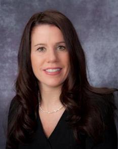
The experts at the UPMC Mercy Burn Center joined the American Burn Association (ABA) in observing National Burn Awareness Week, Feb. 6-12. ABA’s theme this year was “Burning Issues in the Kitchen” as 47% of all home fires are caused by cooking mishaps. According to the American Red Cross, fires break out year-round, but the majority in the United States occur during winter — when conditions for battling them are likely to be at their worst.
“Many of the burns and injuries we see are preventable by using caution and thoughtfulness. Don’t rush or multi-task in the kitchen,” said McGowan Institute for Regenerative Medicine affiliated faculty member Jenny Ziembicki, MD, director of UPMC Mercy Burn Center.
Each year, UPMC Mercy Burn Center treats more than 950 burn patients with varying degrees severity. The UPMC Mercy Burn Center was the first burn center in Pennsylvania, and one of the first in the U.S. As the only burn and level 1 trauma center under one roof in western PA, UPMC Mercy can care for the most critically injured patients that most other hospitals can’t.
Mercy patient and burn survivor Brian Young, 48, was treated at UPMC Mercy last October after suffering burns due to a grease spill. After cooking dinner, Mr. Young left a pot with grease on the stove to cool. However, the electric stove was not turned off. He went to bed and woke up to smoke engulfing his house.
Mr. Young helped his family get outside safely, but while he tried to remove the pot it caught on fire. “I grabbed a pair of gloves from my laundry room. It was so smoky in there and I was choking. I went out the front door and went toward the steps when the grease splashed on me, and I slipped,” he said.
The New Kensington family had suffered a home fire previously and were better prepared to keep the smoke from spreading. “I have a fire extinguisher, but the pot hadn’t combusted yet so I thought I could just remove it.”
Mr. Young sustained burns to his face and his body and was rushed to the hospital. “My wife called the ambulance immediately and they flew me to UPMC Mercy, and the staff took care of me and put me back together,” he shared.
Mr. Young said he believes that fire safety should be taught to everyone especially children, so they become familiar with how dangerous cooking can potentially be and know how to prevent injuries.
Here are some helpful tips to prevent fires and injuries in the kitchen:
- Stay in the kitchen while you are frying, grilling, or broiling food. If you leave, turn off the stove.
- Turn pot or pan handles toward the back of the stove.
- Remain in the home while food is cooking and use a timer to remind you to check on your food.
- After cooking, check the kitchen to make sure all burners and other appliances are turned off.
- NEVER move a pot or carry it outside – the pot is too hot to handle, and the contents may splash, causing a severe burn.
For more than 50 years, the UPMC Mercy Trauma and Burn Center has provided comprehensive care for burn victims of all ages.
Accidental burns are a leading cause of household injuries. If someone in your home sustains a minor burn, remember these important tips:
- Stop the burning process and cover any open areas in a clean cloth.
- Remove any jewelry or clothing in proximity to the burn.
- Do not apply ice and do not break blisters.
The Shift to Using Fewer Opioids

For decades, opioids routinely were prescribed to adults and children for dental procedures, and dentists were among the top prescribers of the drugs after family physicians.
As the opioid crisis has underscored the need to be more judicious when prescribing the drugs, prescriptions for the powerful narcotics have fallen in recent years. Local dentists are increasingly finding alternatives to manage pain — and patients do not seem to mind.
“I found through my own experience treating patients at Children’s Hospital that we could take a non-opioid approach for pretty significant surgeries and patients have a more than adequate experience,” said McGowan Institute for Regenerative Medicine faculty member Bernard Costello, MD, DMD, FACS, Dean of Pitt’s School of Dental Medicine.
Emily Mullin, reporter for the Pittsburgh Post-Gazette, recently spoke with Dr. Costello about why dentists in Western Pa. and across the U.S. are prescribing fewer opioids.
Dr. Costello said he used to prescribe opioids to patients on a “just-in-case” basis so that they would have them on hand if needed. But now his thinking has shifted to a “break glass” approach, in which he only prescribes them after non-opioid medications do not work.
In 2019, Pitt Dental Medicine adopted opioid-free pain management guidelines for most procedures performed in its clinics. The guidelines consider severity, duration and individual risk considerations when prescribing pain medications for patients. In the first year after implementing the plan, opioid prescriptions fell by 54%, according to Dr. Costello. In the second year, they decreased by another 48%.
In December 2021, the dental school pledged that it would incorporate responsible pain management into student and resident training as part of their curriculum. Dr. Costello said training the next generation of dentists to use fewer opioids is key to preventing misuse of the drugs.
Welcome New Faculty Members

The McGowan Institute for Regenerative Medicine welcomes new affiliated faculty members Jarrett Cain, DPM, MSc, FACFAS, Allison Bean, MD, PhD, and Johannes Bonatti, MD, FETCS.
Dr. Jarrett Cain is an Associate Professor of Orthopaedic Surgery, University of Pittsburgh School of Medicine. He completed his Doctorate in Podiatric Medicine at Temple University School of Podiatric Medicine in Philadelphia, PA. Also, he completed post graduate studies in Clinical Epidemiology at the University of Pennsylvania School of Medicine in Philadelphia, and a Master’s of Science in Clinical Research from Drexel University School of Medicine in Philadelphia.
Prior to his appointment in Pittsburgh, Dr. Cain was an Associate Professor, Penn State College of Medicine, Hershey, PA. Dr. Cain holds dual certification by the American Board of Foot & Ankle Surgery in Foot Surgery and Reconstructive Rearfoot and Ankle Surgery.
Dr. Cain’s research interests include disorders of the foot and ankle, diabetic bone healing, flatfoot deformity, and osteoarthritis. He has received various grants and has presented his research at various conferences nationally and internationally. His research team was named a finalist in 2017 for J. Leonard Goldner Award for best basic science research entitled “Effect of Topical Opioid Antagonist on Diabetic Wound Healing in Rat Model.” Dr. Cain also serves on the Editorial Board of the Journal of Foot & Ankle Surgery.
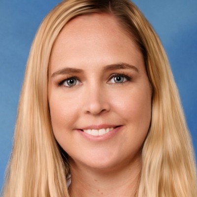 Dr. Allison Bean is an Assistant Professor in the Department of Physical Medicine and Rehabilitation at the University of Pittsburgh School of Medicine.
After receiving a BS in Bioengineering from Rice University, Houston, TX, Dr. Bean continued her studies at the University of Pittsburgh, obtaining a PhD in Bioengineering in 2013 followed by a MD in 2015. Her postgraduate work continued with a preliminary medicine internship at Rutgers-New Jersey Medical School, Newark, NJ, and residency in physical medicine and rehabilitation at Icahn School of Medicine at Mount Sinai, New York, NY. She then completed a sports medicine fellowship in the Department of Physical Medicine and Rehabilitation at the University of Pittsburgh. Dr. Bean is board certified in physical medicine and rehabilitation and sports medicine by the American Board of Physical Medicine and Rehabilitation.
Dr. Bean’s research focuses on understanding the molecular mechanisms of musculoskeletal tissue injury and repair, using this knowledge to guide the development of novel regenerative rehabilitation therapies to improve physical function. She is particularly interested in how sex and aging influence disease and recovery in musculoskeletal disorders such as skeletal muscle and tendon injury. Her work is currently funded by the University of Pittsburgh KL2 scholars program, as well as the Foundation for Physical Medicine and Rehabilitation and the Alliance for Regenerative Rehabilitation Research and Training.
Dr. Bean’s clinical practice focuses on non-surgical treatment of sports injuries, including joint, tendon, and ligament disorders. She specializes in the use of musculoskeletal ultrasound for diagnostic and interventional applications including administration of orthobiologics (e.g., platelet-rich plasma and dextrose prolotherapy).
She is a member of the Association of Academic Physiatrists (Research Committee Member), American Academy of Physical Medicine and Rehabilitation, American Medical Society for Sports Medicine, and the Orthopedic Research Society. Dr. Bean is an Editorial Board Member and Co-Editor Resident Fellow Section for the American Journal of Physical Medicine and Rehabilitation.
 Dr. Johannes Bonatti is an internationally recognized expert in robotic-assisted mitral valve and coronary surgery who is certified by the European Board of Thoracic and Cardiovascular Surgeons. He is a Professor of Cardiothoracic Surgery at the University of Pittsburgh School of Medicine, and the Director of Cardiac Robotic Surgery at the UPMC Heart and Vascular Institute. Dr. Bonatti comes to the UPMC Heart and Vascular Institute from his most recent appointment at the Vienna Health Network in Vienna, Austria. Prior to his work in Austria, Dr. Bonatti was the Chief of the Department of Cardiothoracic Surgery at the Cleveland Clinic Abu Dhabi, a position he held from 2012 to 2015.
He received his medical degree from Leopold Franzens University in Innsbruck, Austria, and completed residencies in Cardiac Surgery, Vascular Surgery, and General Surgery at the University Clinic of Surgery in Innsbruck, followed by internships in Trauma Surgery, Internal Medicine, and Anesthesiology at the General Public Hospital Kitzbühel, Austria.
Dr. Bonatti’s clinical interests include minimally invasive cardiac surgery, robotic cardiac surgery, and vascular biology of coronary artery bypass grafts. He has published more than 250 peer-reviewed articles and performed over 5,000 cardiac surgeries, in addition to being the first physician world-wide to perform a robotic endoscopic quadruple coronary bypass surgery. He has held leadership positions in preeminent surgical societies worldwide.
From a research perspective, one of Dr. Bonatti’s goals is to make cardiac surgical procedures, regardless of the type, as endoscopic as possible. He is also interested in testing emerging robotic surgical technologies and platforms and applying them to ever-wider surgical indications.
Dr. Bonatti directs the clinical and research efforts of the UPMC HVI robotic cardiac surgery program, expanding the institute’s capabilities in robotic cardiac surgery while working to study outcomes and develop new applications, approaches, and indications for robotically assisted procedures. Dr. Bonatti also works in the UPMC robotic surgery simulation lab to develop structured training programs, conduct procedural training for students and surgeons, and further his research aims related to advancing and expanding the discipline of robotic cardiac surgery. Simulation-based training and study on dry-lab and wet-lab models are crucial to developing robotic surgical skills, and UPMC has extensive capabilities on the simulation front for training and research.
As with all robotically assisted surgery, the technology is highly advanced and allows for a fully immersive visual field with better acuity and the ability to manipulate the instrumentation in highly complex ways and more precise control than human hands can accomplish on their own.
Training and mentoring cardiac surgeons on robotic platforms and techniques is a significant part of Dr. Bonatti’s clinical and research work. In the past, he has been the President of the Minimally Invasive Robotics Association (MIRA) and President of ISMICS.
Welcome!!
AWARDS AND RECOGNITION
Dr. Anna Balazs Named Member of the National Academy of Engineering

Less than a year after being elected to the National Academy of Sciences, McGowan Institute for Regenerative Medicine affiliated faculty member and University of Pittsburgh Distinguished Professor Anna Balazs, PhD, was named as one of the newest members of the National Academy of Engineering (NAE). The Academy accepted Dr. Balazs’ membership “in recognition of distinguished contributions to engineering and for creative and imaginative work in predicting the behavior of soft materials that are composed of multiple cooperatively interacting components.”
Dr. Balazs is the first faculty member in the Swanson School of Engineering to hold membership in both the Engineering and Science academies. Dr. Balazs and the 111 new members and 22 international members announced by NAE President John L. Anderson will be inducted at the NAE Annual Meeting this fall, October 2-3 in Washington, DC.
Dr. Balazs’ research focuses on “biomimicry” and the theoretical and computational modeling of polymers. An internationally acclaimed expert in the field, her many accolades also include the first woman to receive the prestigious Polymer Physics Prize from the American Physical Society in 2016. She is also the John A. Swanson Chair of Engineering in the Swanson School’s Department of Chemical and Petroleum Engineering.
“This has been a tremendous year for Anna and long-due recognition of her outstanding career in engineering and chemistry,” noted McGowan Institute faculty member Steven Little, PhD, Department Chair and Distinguished Professor of Chemical Engineering. “Her continued impact in this singular field represents the foundation for soft robotics, advanced materials, and a new interpretation of polymer chemistry. Most importantly, Anna is also a remarkable teacher, mentor, and colleague, especially to our young faculty. All of us share in her excitement today and share a sense of pride in her lifetime accomplishments.”
Election to the National Academy of Engineering is among the highest professional distinctions accorded to an engineer. Academy membership honors those who have made outstanding contributions to “engineering research, practice, or education, including, where appropriate, significant contributions to the engineering literature” and to “the pioneering of new and developing fields of technology, making major advancements in traditional fields of engineering, or developing/implementing innovative approaches to engineering education.”
Dr. Balazs is a fellow of the American Physical Society, the Royal Society of Chemistry, and the Materials Research Society, and received some of the leading awards in her field, including the Royal Society of Chemistry S F Boys – A Rahman Award (2015), the American Chemical Society Langmuir Lecture Award (2014), and the Mines Medal from the South Dakota School of Mines and Technology (2013).
“Anna is truly an exceptional engineer whose passion for polymer chemistry excites and inspires,” said James R. Martin II, PhD, U.S. Steel Dean of Engineering. “With her election to two of our country’s most prestigious national academies, Anna has firmly established herself as an exemplar of innovation and a role model to so many in our Swanson School community. I look forward to soon celebrating her success.”
Congratulations, Dr. Balazs!
Illustration: University of Pittsburgh Swanson School of Engineering.
Dr. Fabrisia Ambrosio Elected to the 2022 Class of the AIMBE College of Fellows

The American Institute for Medical and Biological Engineering (AIMBE) has announced the election of Fabrisia Ambrosio, PhD, MPT, Director of Rehabilitation for UPMC International and an Associate Professor, Physical Medicine & Rehabilitation, University of Pittsburgh, to its College of Fellows. Dr. Ambrosio was nominated, reviewed, and elected by peers and members of the AIMBE College of Fellows for outstanding contributions to the novel field of Regenerative Rehabilitation, integrating applied biophysics and cellular therapeutics to optimize tissue function.
Dr. Ambrosio’s research interests reside at the intersection of physiology and physics and have spanned people to particles.
Dr. Ambrosio recognized the potential of applied biophysics to enhance the efficacy of cellular therapeutics and to provide mechanistic insight into regenerative biology, even beyond the skeletal muscle model that was the focus of her laboratory. Inspired by this potential, she led efforts towards formation of a novel fusion field, ‘Regenerative Rehabilitation’. She has written many articles on the topic, and in 2011, she served as Founding Course Director for the Symposium on Regenerative Rehabilitation. This Symposium has since become an annual event, supported by an international consortium that comprises 17 institutions across North America, Europe, and Asia, of which she is the Founding Director. Dr. Ambrosio is also the lead Principal Investigator for the NIH-funded Alliance for Regenerative Rehabilitation Research & Training, totaling over $10M in funding. The field of Regenerative Rehabilitation has enjoyed steady growth since its inception and has been the topic of multiple journal special issues and DOD funding initiatives. Moreover, Regenerative Rehabilitation was cited in the NIH Research Plan as an example of “actions that will help meet the strategic goals of the NIH institutes and centers that support rehabilitation research.”
Most recently, Dr. Ambrosio has scaled her research to the atomic scale, in which she investigates the contribution of quantum phenomena, such as radical pair reactions, entropy, and entanglement, in dictating stem cell function (i.e., quantum biology). Taken together, Dr. Ambrosio’s record serves as a testament to her unwavering commitment to pioneering novel and convergent research paradigms.
A major focus of Dr. Ambrosio’s programmatic efforts has been to encourage the participation of under-represented minorities in science and technology training programs.
The College of Fellows is comprised of the top two percent of medical and biological engineers in the country. The most accomplished and distinguished engineering and medical school chairs, research directors, professors, innovators, and successful entrepreneurs comprise the College of Fellows. AIMBE Fellows are regularly recognized for their contributions in teaching, research, and innovation. AIMBE Fellows have been awarded the Nobel Prize, the Presidential Medal of Science and the Presidential Medal of Technology and Innovation, and many also are members of the National Academy of Engineering, National Academy of Medicine, and the National Academy of Sciences.
A formal induction ceremony will be held during AIMBE’s 2022 Annual Event on March 25. Dr. Ambrosio will be inducted along with 152 colleagues who make up the AIMBE Fellow Class of 2022.
AIMBE’s mission is to recognize excellence in, and advocate for, the fields of medical and biological engineering to advance society. Since 1991, AIMBE’s College of Fellows has led the way for technological growth and advancement in the fields of medical and biological engineering. AIMBE Fellows have helped revolutionize medicine and related fields to enhance and extend the lives of people all over the world. They have successfully advocated for public policies that have enabled researchers and business-makers to further the interests of engineers, teachers, scientists, clinical practitioners, and ultimately, patients.
AIMBE Fellows are committed to giving back to the fields of medical and biological engineering through advocacy efforts and public policy initiatives that benefit the scientific community, as well as society at large.
Congratulations, Dr. Ambrosio!
Dr. Steve Little Among Newest Class of AAAS Fellows

McGowan Institute for Regenerative Medicine faculty member Steven Little, PhD, Distinguished Professor and Department Chair of Chemical and Petroleum Engineering at the University of Pittsburgh, has been elected Fellow of the American Association for the Advancement of Science (AAAS).
Recognized by AAAS “for remarkable service to charities that advance education in science in impoverished countries and leadership in science internationally,” Dr. Little is one of four Pitt and UPMC faculty named as AAAS Fellows, one of the most distinct honors within the scientific community, dating to 1874.
“Steve’s outstanding career trajectory has been highlighted by the prestigious honors he’s received from his peers and societies like AAAS,” noted Robert Langer, ScD, the David H. Koch Institute Professor and Dr. Little’s PhD advisor at Massachusetts Institute of Technology (MIT). “He is a Renaissance innovator who in his young career has helped to transform drug delivery research and is still poised for greater things.”
At Pitt, Dr. Little also holds the title of William Kepler Whiteford Endowed Professor in the Swanson School of Engineering, as well as secondary appointments in Bioengineering, Pharmaceutical Sciences, Immunology, and Ophthalmology. Dr. Little is among 564 Fellows across a cadre of scientists, engineers and innovators recognized for achievements across disciplines ranging throughout research, teaching, administration, industry, government, and communications.
“I am incredibly humbled by this honor, and grateful for the years of mentorship from Bob Langer as well as the resources I’ve received at the University of Pittsburgh to further my research,” Dr. Little said. “Congratulations to my Pitt colleagues as well as the other Fellows named in this year’s cohort.”
Since receiving his PhD in 2005 – with his thesis winning the AAAS Excellence in Research Award – he has developed numerous new drug formulations including controlled drug release that mimics the body’s own mechanisms of healing and resolving inflammation. Unlike traditional medications that require large doses administered via ingestion, inoculation or intravenously, biomimetic treatments recruit a patient’s own cells to treat disease at the source. In particular, Dr. Little’s research shows potential new applications for glaucoma, gum disease, and even transplant organ rejection.
Congratulations, Dr. Little!
Drs. Antonio D’Amore and Cecelia Yates Elected to The National Academy of Inventors 2022 Class of Senior Members
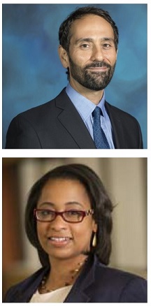
The National Academy of Inventors (NAI) has named 83 of the world’s best emerging academic inventors to its 2022 Class of Senior Members. Two McGowan Institute for Regenerative Medicine affiliated faculty members are a part of this distinguished group. They are:
- Antonio D’Amore, PhD, Research Assistant Professor in the Departments of Surgery and Bioengineering at the University of Pittsburgh
- Cecelia Yates, PhD, Associate Professor in the Department of Health Promotion & Development, School of Nursing at the University of Pittsburgh, with secondary appointments in the Departments of Pathology and Bioengineering
NAI Senior Members are active faculty, scientists, and administrators from NAI Member Institutions who have demonstrated remarkable innovation producing technologies that have brought, or aspire to bring, real impact on the welfare of society. They also have growing success in patents, licensing, and commercialization, while educating and mentoring the next generation of inventors.
This latest class of NAI Senior Members hails from 41 research universities. They are named inventors on over 1093 issued U.S. patents.
This year’s class also reflects NAI’s dedicated efforts to promote diversity and inclusion in its membership, with the addition of 40 outstanding academic female and/or minority inventors. The 2022 new Senior Members will be inducted at the Senior Member Ceremony at the 11th Annual Meeting of the National Academy of Inventors this upcoming June 14-15 in Phoenix, Arizona.
Senior Members are elected annually on National Inventors’ Day (February 11). Nominations for the 2023 Senior Member class will be accepted from October 1 – December 31, 2022.
The National Academy of Inventors is a member organization comprising U.S. and international universities, and governmental and non-profit research institutes, with over 4,000 individual inventor members and Fellows spanning more than 250 institutions worldwide. It was founded in 2010 to recognize and encourage inventors with patents issued from the U.S. Patent and Trademark Office (USPTO), enhance the visibility of academic technology and innovation, encourage the disclosure of intellectual property, educate and mentor innovative students, and translate the inventions of its members to benefit society. The NAI works in partnership with the USPTO and publishes the multidisciplinary journal, Technology and Innovation. Learn more here.
Regenerative Medicine Podcast Update
The Regenerative Medicine Podcasts remain a popular web destination. Informative and entertaining, these are the most recent interviews:
#230 –– Dr. Steven Badylak discusses his research in using artificial intelligence in wound healing.
Visit www.regenerativemedicinetoday.com to keep abreast of the new interviews.
PUBLICATION OF THE MONTH
Author: Bing Han, Maria Giovanna Francipane, Amin Cheikhi, Joycelyn Johnson, Fei Chen, Ruoyu Chen, Eric Lagasse
Title: Fat-associated lymphoid clusters as expandable niches for ectopic liver development
Summary: Background and Aims: Hepatocyte transplantation holds great promise as an alternative approach to whole-organ transplantation. Intraportal and intrasplenic cell infusions are primary hepatocyte transplantation delivery routes for this procedure. However, patients with severe liver diseases often have disrupted liver and spleen architectures, which introduce risks in the engraftment process. We previously demonstrated i.p. injection of hepatocytes as an alternative route of delivery that could benefit this subpopulation of patients, particularly if less invasive and low-risk procedures are required; and we have established that lymph nodes may serve as extrahepatic sites for hepatocyte engraftment. However, whether other niches in the abdominal cavity support the survival and proliferation of the transplanted hepatocytes remains unclear.
Approach and Results: Here, we showed that hepatocytes transplanted by i.p. injection engraft and generate ectopic liver tissues in fat-associated lymphoid clusters (FALCs), which are adipose tissue–embedded, tertiary lymphoid structures localized throughout the peritoneal cavity. The FALC-engrafted hepatocytes formed functional ectopic livers that rescued tyrosinemic mice from liver failure. Consistently, analyses of ectopic and native liver transcriptomes revealed a selective ectopic compensatory gene expression of hepatic function–controlling genes in ectopic livers, implying a regulated functional integration between the two livers. The lack of FALCs in the abdominal cavity of immunodeficient tyrosinemic mice hindered ectopic liver development, whereas the restoration of FALC formation through bone marrow transplantation restored ectopic liver development in these mice. Accordingly, induced abdominal inflammation increased FALC numbers, which improved hepatocyte engraftment and accelerated the recovery of tyrosinemic mice from liver failure.
Conclusions: Abdominal FALCs are essential extrahepatic sites for hepatocyte engraftment after i.p. transplantation and, as such, represent an easy-to-access and expandable niche for ectopic liver regeneration when adequate growth stimulus is present.
Source: Hepatology. 2021 Dec 10. doi: 10.1002/hep.32277. Online ahead of print.
GRANT OF THE MONTH
PI: Kang Kim
Title: Super Resolution Ultrasound Imaging of Vasa Vasorum to Characterize the Progression of Atherosclerotic Plaques and Predict Rupture Vulnerability
Description: Acute coronary syndromes and strokes together constitute a leading cause of morbidity and mortality in the United States and Europe, approximately 80% of which are caused by atherosclerotic plaque (AP) rupture. Over the past decade, extensive efforts have been made to identify a rupture-prone AP. Among others, infiltration of dense neovascularization arising from vasa vasorum (VV) into the AP core plays a critical role in AP rupture. Postmortem studies revealed key involvement of VV in AP. However, a persistent lack of a noninvasive, high-resolution imaging tool to longitudinally assess abnormal microvascular expansion remains a critical barrier to adequate in-vivo investigation on how VV affects AP progression and contributes to eventual rupture. To address this dire unmet need, we propose an innovative transcutaneous super resolution ultrasound (SRU) imaging. The technology development in this project seeks to shift the current US imaging approach in identifying microvessels of AP from “intravascular” to a “fully noninvasive transcutaneous” imaging approach. This is only possible by achieving unprecedented high spatial resolution at large depth, breaking acoustic diffraction limit of the ultrasound frequency that governs spatial resolution. Our group has performed in-depth feasibility studies where SRU imaging successfully identified neomicrovessels in cholesterol-fed rabbit AP, evaluated against µCT and histology. Additionally, areas requiring further technical optimization were identified. Such technology developments and preliminary data thus far rigorously support our overarching hypothesis that enhanced and optimized SRU will accurately stage plaque progression and identify rupture- prone plaques by directly measuring VV changes with exquisite detail. To test the hypothesis, we will use a well-established, clinically relevant cholesterol-fed rabbit AP rupture model, which has shown the most similarity to human plaque pathology including VV neovascularization, to validate the novel SRU system to 1) Successfully quantify changes in vessel density and 2) Identify rupture-prone AP. To achieve these goals, we propose the following specific aims: 1) To develop enhanced SRU at high frequency using a commercial small animal imaging probe 2) To determine if VV changes estimated by SRU correlate with AP progression and are predictive of AP rupture. The immediate outcomes of the proposed work are an affordable noninvasive small animal SRU imaging tool and it’s validation on a clinically relevant rabbit AP model, which also can be used for other important small animal disease models, which are associated with microvessel abnormality such as cancer angiogenesis and kidney diseases to name a few. With proper adaptations into a clinical mid frequency probe and validation in clinical settings in future, this work will lead to our long-term translational goal to integrate SRU in a facile manner into the current clinical standard of carotid duplex sonography that has shown poor specificity to plaque vulnerability. This will help to effectively stratify patients at high risk of strokes and guide adequate intervention/treatment options for stroke prevention, exerting highly influential clinical impact.
Source: National Heart, Lung, and Blood Institute
Term: February 1, 2022 – January 31, 2026
Amount: $694,630 (one year)
