June 2015 | VOL. 14, NO. 6 | www.McGowan.pitt.edu
Computer Simulation Accurately Replicated Real-Life Trauma Outcomes
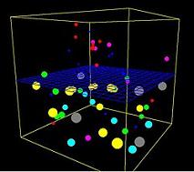 A computer simulation, or in silico model, of the body’s inflammatory response to traumatic injury accurately replicated known individual outcomes and predicted population results counter to expectations, according to a study recently published in Science Translational Medicine by a University of Pittsburgh research team which included McGowan Institute for Regenerative Medicine affiliated faculty members—Yoram Vodovotz, PhD, professor of surgery and director of the Center for Inflammation and Regenerative Modeling at the University of Pittsburgh School of Medicine, Timothy Billiar, MD, George Vance Foster professor and chair, Pitt Department of Surgery, Ruben Zamora, PhD, research associate professor at the Pitt’s Department of Surgery, and Gregory Constantine, PhD, professor of mathematics and statistics at Pitt.
Traumatic injury is a major health care problem worldwide. Trauma induces acute inflammation in the body with the recruitment of many kinds of cells and molecular factors that are crucial for tissue survival, explained senior investigator Dr. Vodovotz. But if inappropriately sustained, the inflammatory response can compromise healthy tissues and organs.
“Thanks to life-saving surgery and extensive supportive care, most patients who require trauma care are now highly likely to survive,” Dr. Vodovotz said. “But along the way, they may experience a variety of complications, such as multiple organ failure, that are difficult to predict an initial assessment. Our current challenge is to identify which patients are vulnerable to certain problems so that we can better implement surveillance and prevention strategies and use resources more effectively.”
Building from a model developed for swine, the research team examined blood samples from 33 survivors of car or motorcycle accidents or falls for multiple markers of inflammation, including interleukin-6 (IL-6), and segregated the patients into one of three (low to high) categories of trauma severity. They were able to validate model predictions regarding hospital length of stay in a separate group of nearly 150 trauma patients. They then generated a set of 10,000 “virtual patients” with similar injuries and found the model could replicate outcomes in individuals, such as length of stay and degree of multi-organ dysfunction. Intriguingly, the in silico model also predicted a 3.5 percent death rate, comparable to published values and to the Pitt group’s own observations, even though the model did not include patients who didn’t survive their injuries.
The in silico model predicted that, on an individual basis, virtual patients who made more IL-6 in response to trauma were less likely to survive. But, as the model predicted, that was not true at the population level: Among nearly 100 real patients whose genetic predisposition to make more or less amounts of IL-6 had been determined, there was little difference in survival between high- and low-IL-6 producers.
“These findings demonstrate the limitations of extrapolating from single mechanisms to outcomes in individuals and populations, which is the typical paradigm used to identify potential treatments,” Dr. Vodovotz said. “Instead, dynamic computational models like ours that simulate multiple factors that interact with each other in complex diseases could be a more efficient and accurate way of predicting outcomes for both individuals and populations. Then we can pursue those avenues that have the greatest likelihood of success in clinical trials.”
“The potential impact of this work is high because clinical trials are difficult and expensive to carry out, and usually can test only a single dose of a drug,” noted co-investigator Dr. Billiar. “Determining the best dose, timing, and biomarkers that would characterize patients likely to respond well to therapy is a major thrust of the pharmaceutical and biotechnology companies. This approach could help tailor treatments.”
Illustration: Wikipedia.
RESOURCES AT THE MCGOWAN INSTITUTE
July Special at the Histology Lab
Mucins are a family of high molecular weight, heavily glycosylated proteins (glycoconjugates) produced by epithelial tissues in most metazoans. Mucins’ key characteristic is their ability to form gels; therefore they are a key component in most gel-like secretions, serving functions from lubrication to cell signalling to forming chemical barriers. They often take an inhibitory role. Some mucins are associated with controlling mineralization, and bone formation in vertebrates. They bind to pathogens as part of the immune system. Overexpression of the mucin proteins, especially MUC1, is associated with many types of cancer.
Alcian Blue/PAS
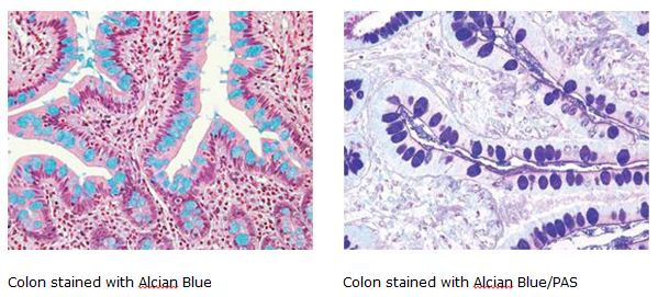
You’ll receive 30% off your Alcian Blue or PAS staining or both when you mention this article any time in July. As always, you will receive the highest quality histology in the quickest turn-around time. Contact Lori at the McGowan Core Histology Lab and ask about our Staining specials. Email perezl@upmc.edu or call 412-624-5265.
Did you know the more samples you submit to the histology lab the less you pay per sample? Contact Lori to find out how!
Flow Cytometry at the McGowan Institute
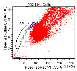 Do you need to:
- Purify samples?
- Characterize complex samples?
- Define a rare subpopulation?
- Examine cellular function?
- Study proliferation or apoptosis?
- Design a flow experiment and need help?
Check out our website or contact Lynda for more information.
UPCOMING EVENTS
Fourth Annual Regenerative Rehabilitation Symposium
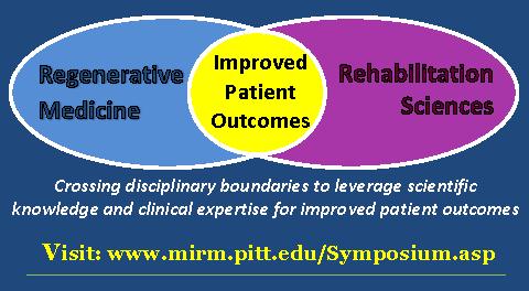 The annual Regenerative Rehabilitation Symposia series is a unique opportunity for students, researchers, and clinicians working in the interrelated fields of regenerative medicine and rehabilitation to meet, exchange ideas, and generate new collaborations and clinical research questions. Jointly organized by the University of Pittsburgh Rehabilitation Institute, the School of Health and Rehabilitation Sciences at the University of Pittsburgh, the McGowan Institute for Regenerative Medicine and the Rehabilitation Research and Development Center of Excellence at the Veterans Affairs Palo Alto Health Care System, the Fourth Annual Symposium on Regenerative Rehabilitation will be held on September 24-26, 2015 in Rochester, MN, hosted by the Mayo Clinic.
For more information on this event, please contact Katy Wharton at: rehabmtg@pitt.edu or whartonkm@upmc.edu or call 412-624-5293.
SCIENTIFIC ADVANCES
Statistical Approach Able To Pinpoint Real from Artifact Alerts
 As reported by Michael Vlessides of Anesthesia News, using a statistical approach known as Random Forest modeling, real and artifact vital sign events from continuous monitoring data have been distinguished. The approach is an important step toward reducing the alarm fatigue that plagues so many health care practitioners.
“We’ve had a long history of using machine learning to assess the instability of patients in the hospital,” said McGowan Institute for Regenerative Medicine affiliated faculty member Michael Pinsky, MD, professor of critical care medicine, bioengineering, anesthesiology, cardiovascular diseases, and clinical/translational services at the University of Pittsburgh School of Medicine. “In one such study, we analyzed noninvasive vital signs such that alerts would go off if the monitor value was outside the normal range,” he said. “And using an artificial neural net, we were able to identify stable from unstable, good from bad. This current study is an extension of that work.”
In the study which also included the efforts of McGowan Institute for Regenerative Medicine affiliated faculty member Gilles Clermont, MD, professor of critical care medicine, industrial engineering, and mathematics at the University of Pittsburgh, noninvasive monitoring data were recorded for patients in the institution’s 24-bed step-down unit over a period of 8 weeks. The data included heart rate (HR), respiratory rate (RR), blood pressure (BP), and peripheral oximetry. Deviations of vital signs beyond stability thresholds that persisted for 80% of a 5-minute moving window comprised important events. These stability thresholds were defined as HR 40 to 140 beats per minute; RR eight to 36 breaths per minute; systolic BP 80 to 200 mm Hg; diastolic BP less than 110 mm Hg; peripheral oxygen saturation (SpO2) greater than 85%.
The researchers, reporting at the 44th Critical Care Congress of the Society of Critical Care Medicine (abstract 42), found that of 1,582 events, 631 were labeled by consensus of four expert clinicians as either real alerts, artifacts, or “unable to classify;” another 795 were labeled as “unseen.” Random Forest models, which use an ensemble of algorithms that together provide increasing predictive accuracy, were then applied to the 631 labeled events; the models were trained to differentiate real events from artifacts, and then were cross-validated to mitigate overfitting. The resulting model was then applied to the 795 unseen events, which were reviewed by the experts for external validation of Random Forest events.
“We found that in this specific example, Random Forest was the most predictive of all the machine learning approaches,” Dr. Pinsky said in an interview with Anesthesiology News. “This is where you need a marriage of clinicians and machine learning science. Neither one on its own would be able to come up with anything of relevance; it has to be a union.”
“So our new approach allows us, with a high degree of certainty, to identify almost all alerts that are real or artifact,” Dr. Pinsky said. “This turned out to be the real excitement at the meeting, for which we won the award as best abstract. Clearly, this is really important for the bedside practitioners.”
Designing 3D-Printed Materials to Help the Body Heal Itself
 In the laboratory of McGowan Institute for Regenerative Medicine affiliated faculty member Prashant Kumta, PhD, the Edward R. Weidlein chair professor at the University of Pittsburgh Swanson School of Engineering and School of Dental Medicine and professor in the Departments of BioEngineering, Chemical and Petroleum Engineering, Mechanical Engineering and Materials Science, and Oral Biology, one of the visions is to revolutionize metallic biomaterials and technologies to create life changing devices. This will lead to engineered systems that will interface with the human body to prolong and improve quality of life, specifically with craniofacial and orthopedic applications. Dr. Kumta and his team’s goal is to engineer logical and clinically relevant options that could regenerate mineralized tissue (bone/tooth) formation utilizing a unique combination of evolutionary load-bearing biodegradable materials (metals), growth factors, and cell therapy.
Recently, Rosanne Skirble of Voice of America News reported (video here) that Dr. Kumta and his team of graduate students are designing 3D-printed materials that are a match for the patient’s body, and are absorbed or excreted as new tissue grows and the wound heals. In the laboratory, Dr. Kumta’s team has developed both magnesium and iron alloys to use as the materials’ base. He calls magnesium — a mineral needed for more than 300 biochemical reactions in the body — “a perfect fit” for the technique.
“It has the mechanical characteristics that meet natural bone, both from the strength [and] the toughness as well as the density. It has the perfect density that will match with natural bone,” he said.
Dr. Kumta’s team is also working with a novel formulation of calcium phosphate putty that can be injected to fill spaces between fractured bones. A 3D-printed biodegradable plate or screw would hold that filler in place.
“The fixation plate will provide the mechanical strength needed to carry the load, and the bone-wide filler would help provide the healing and the bone formation,” he said.
Pre-clinical trials are now underway in animals. Dr. Kumta said the work is revolutionary, offering the prospect of a better outcome for the patient.
“Rather than implanting a screw or plate or joint,” he said, “doctors could give the body’s own regenerative ability a more effective method to heal itself.”
Regenerating Diminished Skeletal Muscle Tissue
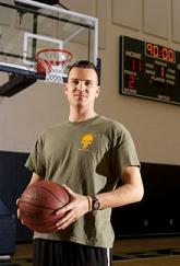 Regenerative medicine uses clinical procedures to repair or replace damaged or diseased tissue and organs vs. some of the traditional therapies that are designed to just treat the symptoms. In Pitt Magazine, Holden Slattery recently reported on the progress being made by patients enrolled in a University of Pittsburgh-University of Pittsburgh Medical Center study that tests a new approach to significant muscle loss.
Co-led by McGowan Institute for Regenerative Medicine faculty members Stephen Badylak, DVM, PhD, MD, Professor in the Department of Surgery, a Deputy Director of the McGowan Institute, and Director of the Center for Pre-Clinical Tissue Engineering within the Institute, and J. Peter Rubin, MD, Chair of the Department of Plastic Surgery, UPMC Endowed Professor of Plastic Surgery, and Professor of Bioengineering at the University of Pittsburgh, the research team uses extracellular matrix (ECM) from a pig’s bladder to regenerate diminished skeletal muscle. ECM is the remaining tissue after all the cells have been removed from the pig’s bladder. Dr. Badylak has been researching the effects of ECM since 1987. This study is the first to utilize ECM to replace and regenerate muscle tissue.
In each of the study’s five surgeries, scar tissue from the damaged leg muscles was removed and ECM was placed near healthy muscle. Within months, mature skeletal muscle cells emerged at the site of the ECM placement, along with other cells that appeared to be actively regenerating into skeletal muscle cells. Eventually, the ECM dissolved leaving only healthy muscle tissue.
Three of the five patients have shown a 20% or greater improvement in strength of the injured leg. The results haven’t only shown dramatic improvement on physical tests. The participants are now engaging in a range of activities including running, bicycling, playing soccer, and even skiing—activities they couldn’t enjoy before their surgery.
“We love science, and we’re very interesting in these stem cells,” Dr. Badylak says. “But at the end of the day the story is incomplete without the impact on patients’ daily activities.”
Illustration: Regenerative medicine patient Sgt. Ronald Strang. – Pitt Magazine.
Stem Cell and Epigenetic Strategies for Retinal Repair
 McGowan Institute for Regenerative Medicine affiliated faculty member Igor Nasonkin, PhD, has recently learned how to grow a 3D layer of early human retinal cells in the laboratory, with hopes of one day implanting them in people with damaged retinas. Dr. Nasonkin, who has a doctorate in human genetics and is director of the University of Pittburgh’s Retinal Repair Laboratory and the assistant director of the Louis J. Fox Center for Vision Restoration of the University of Pittsburgh Medical Center and University of Pittsburgh, doesn’t want to pinpoint when his techniques might be tried in human patients, but said other kinds of human tissue transplants in the eye have paved the way for his group’s work. His research was recently highlighted by Mark Roth of the Pittsburgh Post-Gazette.
“It looks like if you push cells to a certain stage,” Dr. Nasonkin said, “they can self-assemble as long as you feed them the right nutrition.” And that means scientists may soon be able to create a complete “retinal patch” for people with retinitis pigmentosa or other diseases.
Photoreceptor death in retinal and macular degenerative diseases is a leading cause of inherited vision loss in developed countries. Vision loss may be devastating and urgently requires new ideas, approaches, and paradigms on the molecular level, to alleviate this condition. Novel therapeutic strategies have recently emerged, from mechanical to cell-based, to repair neural circuits affected by photoreceptor (PR) cell loss. Trophic factor delivery to extend the life of dying PRs has been pursued in experimental animals, and in some instances, in the clinic. Gene therapy approaches have been applied successfully in one type of Leber congenital amaurosis (LCA). Retinal implants designed to capture photons and transmit the electric signals to ganglion cells have been used in the clinic with some promising outcomes.
The transplantation of retinal cells into mammalian retina produces variable outcomes and success, yet seems very promising due to some rare successful experiments demonstrating the efficient integration of retinal cells into the recipient retina. A greater understanding of biology and improvements in methodology are required before such protocols may be introduced into the clinic. The major obstacles to retinal cell replacement are the elusive molecular signature of the “transplantable” retinal progenitor, the specific synaptic integration of such cells into preexisting neural retinal circuitry of the host, and the ability of grafted retinal cells to migrate through the impermeable outer limiting membrane and position themselves into the required retinal niche (such as photoreceptor layer).
A combination of retinal stem cell grafting and trophic factor delivery has recently emerged as a fruitful direction to alleviate vision loss by slowing down the death of retinal cells, especially photoreceptors. Such direction may not necessarily require the integration of transplanted cells into specific retinal layer, yet may produce long lasting positive therapeutic effect, and should be definitely explored further. Induction of the endogenous regeneration in mammalian CNS, including retina, is still an unattainable goal of therapies aimed at repairing age-related vision loss and retinal trauma. However, some lower species, such as fish, amphibians, and to some extent, birds, have active regenerative mechanisms in the eye, which last throughout their lives.
Regeneration requires the endogenous retinal cells (either dormant stem cells or postmitotic mature cells in retina) to reenter the mitotic cycle, differentiate into the cell type needed for retinal repair, and replenish the lost cells. Understanding the molecular mechanisms governing the developmental plasticity of a retinal cell is the key to be able to induce mature retinal cells to revert to immature progenitors. Epigenetic mechanisms are one likely avenue, which should be explored to change the plasticity of retinal cells and enhance the endogenous regeneration in the retina.
Eventually, Dr. Nasonkin envisions a retinal eye bank derived from adult stem cells that will contain enough different types of human tissue to be compatible with most people’s immune systems.
Lower Birth Weight Associated with Proximity of Mother’s Home to Gas Wells
 Pregnant women living close to a high density of natural gas wells drilled with hydraulic fracturing were more likely to have babies with lower birth weights than women living farther from such wells, according to a University of Pittsburgh Graduate School of Public Health analysis of southwestern Pennsylvania birth records.
The finding does not prove that the proximity to the wells caused the lower birth weights, but it is a concerning association that warrants further investigation, the researchers concluded. The study was funded by The Heinz Endowments and published in PLOS ONE.
“Our work is a first for our region and supports previous research linking unconventional gas development and adverse health outcomes,” said co-author McGowan Institute for Regenerative Medicine affiliated faculty member Bruce Pitt, PhD, chair of Pitt Public Health’s Department of Environmental and Occupational Health. “These findings cannot be ignored. There is a clear need for studies in larger populations with better estimates of exposure and more in-depth medical records.”
Unconventional gas development includes horizontal drilling and high volume hydraulic fracturing, known as “fracking.” It allows access to large amounts of natural gas trapped in shale deposits. Prior to 2007, only 44 wells were known to be drilled in Pennsylvania’s Marcellus Shale with such technology. From 2007 to 2010, that expanded to 2,864 wells.
The Pitt Public Health research team cross-referenced birth outcomes for 15,451 babies born in Washington, Westmoreland, and Butler counties from 2007 through 2010 with the proximity of the mother’s home to wells drilled using unconventional gas development. They divided the data into four groups, depending on the number and proximity of wells within a 10-mile radius of the mothers’ homes.
Mothers whose homes fell in the top group for proximity to a high density of such wells were 34 percent more likely to have babies who were “small for gestational age” than mothers whose homes fell in the bottom 25 percent. Small for gestational age refers to babies whose birth weight ranks them below the smallest 10 percent when compared to their peers.
“Developing fetuses are particularly sensitive to the effects of environmental pollutants,” said Dr. Pitt. “We know that fine particulate air pollution, exposure to heavy metals and benzene, and maternal stress all are associated with lower birth weight.”
In southwestern Pennsylvania, the waste fluids produced through hydrofracturing, called “flowback,” can contain benzene. Unconventional gas development also creates an opportunity for air pollution through flaring of methane gas at the well heads and controlled burning of natural gas that releases volatile organic compounds, including benzene, toluene, ethylbenzene, and xylene. Increased truck traffic and diesel-operated compressors also can contribute to air and noise pollution.
“It is important to stress that our study does not say that these pollutants caused the lower birth weights,” said Dr. Pitt. “Unconventional gas development is dynamic and varies from site to site, changing the potential for human exposure. To draw firm conclusions, we need studies that thoroughly assess the exposure of a very large number of pregnant women to not just the gas wells, but other potential pollutants.”
Shaina L. Stacy, PhD, a recent graduate of Pitt Public Health, is lead author on this research, and Evelyn Talbott, DrPH, epidemiology professor at the school, is senior author. Additional authors are LuAnn L. Brink, PhD, and Bernard D. Goldstein, MD, both of Pitt Public Health; and Jacob C. Larkin, MD, and Yoel Sadovsky, MD, both of Magee-Womens Research Institute and Pitt School of Medicine.
AWARDS AND RECOGNITION
Dr. George Michalopoulos Inducted Into AAP
 McGowan Institute for Regenerative Medicine affiliated faculty member George Michalopoulos, MD, PhD, has been inducted into the Association of American Physicians (AAP), a nonprofit, professional organization founded in 1885 for the “advancement of scientific and practical medicine.”
Dr. Michalopoulos is the Maud L. Menten Professor of Experimental Pathology and chair of the Department of Pathology. His research focuses on new therapies for liver fibrosis and the disease mechanisms in alpha-1 antitrypsin deficiency.
Dr. Michalopoulos received his medical doctoral degree at Athens University School of Medicine, Athens, Greece, in 1969. A residency in Anatomic Pathology and a PhD study in Oncology were completed at the Wisconsin Medical Center in Madison in 1977. Dr. Michalopoulos moved to Duke University as an Assistant Professor in 1977 and stayed at Duke University until 1991. He then moved to Pittsburgh and joined the University faculty in April of 1991. In addition to his current appointment, Dr. Michalopoulos also served as Associate Vice Chancellor for Health Sciences and Interim Dean of the School of Medicine from November of 1995-1998. In 2012, he was honored with the title, University of Pittsburgh Distinguished Professor.
Election to AAP is an honor extended to individuals with outstanding credentials in biomedical science and/or translational biomedical research and is limited to 60 inductees per year. An association of the country’s most accomplished physician-scientists, AAP serves as a forum to create and disseminate knowledge and as a source of inspiring role models for upcoming generations of physicians and medical scientists.
Congratulations, Dr. Michalopoulos!
Badylak Lab Student a Fulbright Scholar
 Catalina Pineda Molina is a doctoral student in the Bioengineering Department at the University of Pittsburgh and works in the laboratory of McGowan Institute for Regenerative Medicine deputy director Stephen Badylak, DVM, PhD, MD. Her research interests are focused on the immune-modulatory effects of adipose derived stem cells used in combination with extracellular matrix scaffolds for regenerative medicine applications.
As reported by Christina Rouvalis, Pitt Magazine, Ms. Molina is a Fulbright Scholar striving to help humanity from a scientific angle. She does research to create new devices to treat disabling conditions such as osteoarthritis, diabetes, and loss of tissue due to trauma. She also wants to advance biotechnological research in Columbia (her home country), where the field is still in its infancy.
The Fulbright Program is a program of highly competitive, merit-based grants for international educational exchange for students, scholars, teachers, professionals, scientists, and artists, founded by United States Senator J. William Fulbright in 1946. Under the Fulbright Program, competitively selected U.S. citizens may become eligible for scholarships to study, conduct research, or exercise their talents abroad; and citizens of other countries may qualify to do the same in the United States. The Fulbright Program is one of the most prestigious awards programs worldwide, operating in over 155 countries. The program was established to increase mutual understanding between the people of the United States and other countries through the exchange of persons, knowledge, and skills. The Fulbright Program provides 8,000 grants annually to undertake graduate study, advanced research, university lecturing, and classroom teaching. As of 2013, more than 325,400 persons—122,800 from the United States and 202,600 from other countries—have participated in the program since it began.
 Regenerative Medicine Podcast Update
The Regenerative Medicine Podcasts remain a popular web destination. Informative and entertaining, these are the most recent interviews:
#148 – Dr. Kirk Conrad is a Professor in the Departments of Physiology and Functional Genomics, and of Obstetrics and Gynecology at the University of Florida College of Medicine. Dr. Conrad discusses his research in the mechanisms underlying the massive systemic maternal vasodilation and increased arterial compliance that transpire during normal pregnancy.
Visit www.regenerativemedicinetoday.com to keep abreast of the new interviews.
Picture of the Month
The Picture of the Month is a compliment to the longstanding features Grant of the Month and Publication of the Month. Each of these features highlights the achievements of McGowan affiliated faculty and their trainees. As we have always welcomed suggestions for grants and publications, please also consider submitting images that can highlight your pioneering work.
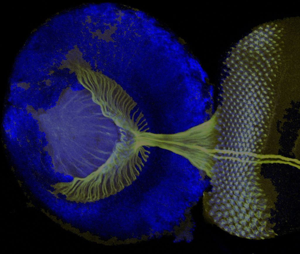
Developing retina and brain of the Drosophila pre-pupal stage animal is viewed as a whole-mount tissue stained with nuclear stain DAPI (Blue) and MAb22C10 (green) that highlights the growing axons of photoreceptor neurons as they extend into the brain were they fan out in the brain to build the retinotopic map. Confocal serial plane reconstruction. By John A. Pollock.
