Artificial Cells to Study Effects of Molecular Crowding on Gene Expression Developed
 The interior of a living cell is a crowded place, with proteins and other macromolecules packed tightly together. A team of scientists at Carnegie Mellon University (CMU), including McGowan Institute for Regenerative Medicine affiliated faculty member Philip LeDuc, PhD, CMU professor of mechanical engineering and biological sciences, has approximated this molecular crowding in an artificial cellular system and found that tight quarters help the process of gene expression, especially when other conditions are less than ideal.
As the researchers report in an advance online publication by the journal Nature Nanotechnology, these findings may help explain how cells have adapted to the phenomenon of molecular crowding, which has been preserved through evolution. And this understanding may guide synthetic biologists as they develop artificial cells that might someday be used for drug delivery, biofuel production, and biosensors.
Most studies of synthetic biological systems today employ solution-based chemistry, which does not involve molecular crowding. The findings of the CMU study and the lessons of evolution suggest that bioengineers will need to build crowding into artificial cells if synthetic genetic circuits are to function as they would in real cells.
The research team, which also included Russell Schwartz, professor of biological sciences; Marcel Bruchez, associate professor of biological sciences and chemistry; and Saumya Saurabh, a doctoral student in chemistry, developed its artificial cellular system using molecular components from bacteriophage T7, a virus that infects bacteria that is often used as a model in synthetic biology.
To mimic the crowded intracellular environment, the researchers used various amounts of inert polymers to gauge the effects of different density levels.
Crowding in a cell isn’t so different from a crowd of people, Tan said. If only a few people are in a room, it’s easy for people to mingle or even to become isolated. But in a crowded room where it’s hard to move around, individuals often will tend to stay close to each other for extended periods. The same thing happens in a cell. If the intracellular space is crowded, binding between molecules increases.
Notably, the researchers found that the dense environments also made gene transcription less sensitive to environmental changes. When the researchers altered concentrations of magnesium, ammonium, and spermidine — chemicals that modulate the stability and binding of macromolecules — they found higher perturbations of gene expression in low-density environments than in high-density environments.
“Artificial cellular systems have tremendous potential for applications in drug delivery, bioremediation, and cellular computing,” Tan said. “Our findings underscore how scientists could harness functioning mechanisms of natural cells to their advantage to control these synthetic cellular systems, as well as in hybrid systems that combine synthetic materials and natural cells.”
Save the Date: Distinguished Lecture October 10th
 The McGowan Institute’s Fall Distinguished Lecturer is Glenn Prestwich, PhD, Presidential Professor of Medicinal Chemistry and Research Professor of Biochemistry, University of Utah. He is also the Director for two Utah Centers of Excellence for technology commercialization: Center for Cell Signaling, and the Center for Therapeutic Biomaterials.
The lecture is scheduled for October 10, 2013 at 4:00 pm at the University Club, Ballroom B. A reception with light refreshments will follow.
Dr. Prestwich graduated with honors with a degree in Chemistry from the California Institute of Technology in 1970. He then earned a PhD in Chemistry from Stanford University. He did two NIH postdoctoral fellowships: at Cornell University and then at the International Centre for Insect Physiology and Ecology in Nairobi, Kenya.
SCIENTIFIC ADVANCES
Practice Makes the Brain’s Motor Cortex More Efficient
 Not only does practice make perfect, it also makes for more efficient generation of neuronal activity in the primary motor cortex, the area of the brain that plans and executes movement, according to McGowan Institute for Regenerative Medicine affiliated faculty member Peter Strick, PhD, and researchers from the University of Pittsburgh School of Medicine. Their findings, published online in Nature Neuroscience, showed that practice leads to decreased metabolic activity for internally generated movements, but not for visually guided motor tasks, and suggest the motor cortex is “plastic” and a potential site for the storage of motor skills.
The hand area of the primary motor cortex is known to be larger among professional pianists than in amateur ones. This observation has suggested that extensive practice and the development of expert performance induces changes in the primary motor cortex, said senior investigator Dr. Strick, Distinguished Professor and chair, Department of Neurobiology, Pitt School of Medicine.
Prior imaging studies have shown that markers of synaptic activity, meaning the input signals to neurons, decrease in the primary motor cortex as repeated actions become routine and an individual develops expertise at a motor skill. The researchers found that markers of synaptic activity also display a marked decrease in monkeys trained to perform sequences of movements that are guided from memory — an internally generated task — rather than from vision. They wondered whether the change in synaptic activity indicated that neuron firing also declined. To examine this issue they recorded neuron activity and sampled metabolic activity, a measure of synaptic activity in the same animals.
All the monkeys were trained on two tasks and were rewarded when they reached out to touch an object in front of them. In the visually guided task, a visual target showed the monkeys where to reach and the end point was randomly switched from trial to trial. In the internally generated task the monkeys were trained to perform short sequences of movements without visual cues. They practiced the sequences until they achieved a level of skill comparable to an expert typist.
The researchers found neuron activity was comparable between monkeys that performed visually guided and internally generated tasks. However, metabolic activity was high for the visually guided task, but only modest during the internally generated task.
“This tells us that practicing a skilled movement and the development of expertise leads to more efficient generation of neuron activity in the primary motor cortex to produce the movement. The increase in efficiency could be created by a number of factors such as more effective synapses, greater synchrony in inputs and more finely tuned inputs,” Dr. Strick noted. “What is really important is that our results indicate that practice changes the primary motor cortex so that it can become an important substrate for the storage of motor skills. Thus, the motor cortex is adaptable, or plastic.”
Premature Aging of Immune Cells Present in Joints of Kids with Chronic Arthritis
 The joints of children with the most common form of chronic inflammatory arthritis contain immune cells that resemble those of 90-year-olds, according to a new study led by researchers at Children’s Hospital of Pittsburgh of UPMC and the University of Pittsburgh School of Medicine. The findings, published recently in Arthritis and Rheumatism, suggest that innovative treatment approaches could aim to prevent premature aging of immune cells.
Juvenile idiopathic arthritis, or JIA, is the most prevalent rheumatic condition in the world and affects 1 of every 1,000 children in the U.S., said senior researcher and McGowan Institute for Regenerative Medicine affiliated faculty member Abbe de Vallejo, PhD, associate professor of pediatrics and immunology, Pitt School of Medicine. It usually starts with a swollen ankle, knee, or wrist that parents often assume is due to a minor injury sustained while playing.
“Untreated JIA has devastating consequences,” Dr. de Vallejo said. “It can slow growth and, in extreme cases, the child can be physically disfigured. It’s a degenerative disease that eats up the joints.”
Doctors have long thought of JIA as an autoimmune disease, meaning the body attacks itself. But previous studies by Dr. de Vallejo of young adults with rheumatoid arthritis indicated that a certain population of cells present in the joint synovial fluid and blood displayed telltale signs of abnormal cell division and premature aging. His current team at Children’s wanted to see if that was true in pediatric arthritis.
They examined immune cells called T-cells in the synovial fluid and blood from 98 children ages 1 to 17 and known to have JIA, as well as 46 blood samples from children who didn’t have the disease. T-cells are the army of immune cells that eradicate infection, tumors, and other dangerous agents to which people may be exposed.
The research team found about one-third of the T-cells of children with JIA had shortened telomeres and had reduced, or in some cases lost, the capacity to proliferate. Telomeres are the ends of chromosomes that don’t code for proteins and, because they are not fully copied by enzyme mechanisms, are trimmed slightly during each DNA replication cycle. It is thought that aging occurs when the telomeres become too short for DNA replication and cell division to proceed normally.
“The T-cells of the children with JIA had very short telomeres, about the length we see in a 90-year-old or a young adult with rheumatoid arthritis. Those same T-cells express unusually high levels of several classic protein markers of cell aging and exhaustion,” Dr. de Vallejo said. “These kids haven’t lived long enough to have cells that look that old. This is the first indication that premature aging is occurring in this childhood condition.”
In addition, the T-cells had become dysregulated, and their immune activity could be stimulated through atypical cell surface receptors. Much more must be learned about the unusual cells and about genetic mechanisms that might contribute to the development of JIA, Dr. de Vallejo said, but these findings could point the way to new therapies.
“JIA is typically treated with broad-spectrum drugs such as steroids and biologics that essentially paralyze the entire immune system, but only a third of the cells are affected and their abnormality seems to be premature aging, rather than autoimmune activity,” he noted. “This study suggests cell-targeted treatments could be developed to prevent this premature immune aging.”
Light That Moves and Molds Gels
 Some animals—like the octopus and cuttlefish—transform their shape based on environment, fending off attackers or threats in the wild. For decades, researchers have worked toward mimicking similar biological responses in non-living organisms, as it would have significant implications in the medical arena.
Now, a team of researchers at the University of Pittsburgh including McGowan Institute for Regenerative Medicine affiliated faculty member Anna Balazs, PhD, has demonstrated such a biomimetic response using hydrogels—a material that constitutes most contact lenses and microfluidic or fluid-controlled technologies. Their study, published in Advanced Functional Materials, is the first to show that these gels can be both reconfigured and controlled by light, undergoing self-sustained motion—a uniquely biomimetic behavior.
“Imagine an apartment with a particular arrangement of rooms all in one location,” said lead author Dr. Balazs, Pitt Distinguished Professor of Chemical and Petroleum Engineering in the Swanson School of Engineering. “Now, consider the possibility of being able to shine a particular configuration of lights on this structure and thereby completely changing not only the entire layout, but also the location of the apartment. This is what we’ve demonstrated with hydrogels.”
Together with Olga Kuksenok, PhD, research associate professor in the Swanson School, Dr. Balazs experimented with a newer type of hydrogel containing spirobenzopyran molecules. Such materials had been previously shown to form distinct 2-D patterns on initially flat surfaces when introduced to varying displays of light and are hydrophilic (“liking” water) in the dark but become hydrophobic (“disliking” water) under blue light illumination. Therefore, Drs. Balazs and Kuksenok anticipated that light could be a useful stimulus for tailoring the gel’s shape.
Using computer modeling, the Pitt team demonstrated that the gels “ran away” when exposed to the light, exhibiting direct, sustained motion. The team also factored in heat—combining the light and local variations in temperature to further control the samples’ motions. Controlling a material with light and temperature could be applicable, Dr. Balazs said, in terms of regulating the movement of a microscopic “conveyor belt” or “elevator” in a microfluidic device.
“This theoretical modeling points toward a new way of configuring the gels into any shape, while simultaneously driving the gels to move due to the presence of light,” said Dr. Kuksenok.
“Consider, for example, that you could take one sheet of hydrogel and, with the appropriate use of light, fashion it into a lens-shaped object, which could be used in optical applications,” added Dr. Balazs.
The team also demonstrated that the gels could undergo dynamic reconfiguration, meaning that, with a different combination of lights, the gel could be used for another purpose. Reconfigurable systems are particularly useful because they are reusable, leading to a significant reduction in cost.
“You don’t need to construct a new device for every new application,” said Dr. Balazs. “By swiping light over the system in different directions, you can further control the movements of a system, further regulating the flow of materials.”
Dr. Balazs said this type of dynamic reconfiguration in response to external cues is particularly advantageous in the realm of functional materials. Such processes, she said, would have a dramatic effect on manufacturing and sustainability, since the same sample could be used and reused for multiple applications.
The team will now study the effect of embedding microscopic fibers into the gel to further control the shape and response of the material to other stimuli.
Click here to see Dr. Balazs give a web seminar about these reconfigurable gels.
Coulter Foundation Translational Research Partnership Program Invests in 3 Medical Technologies of McGowan Affiliated Faculty
 Fighting infection post-surgery with an antibiotic gel, developing a meniscus implant for temporomandibular joint (TMJ) patients, and attacking skin cancer with a microneedle bandage were three of the latest four innovative medical technologies selected for funding through the Wallace H. Coulter Translational Research Partners II (TPII) Program (Coulter Program) this year at the University of Pittsburgh.
“We are excited because these technologies have already generated promising early animal test results, are being patent-protected, have significant clinical and commercial potential, and would benefit from a business focus and a financial “push” toward commercialization,” explained Pratap Khanwilkar, PhD, MBA, professor of bioengineering at the Swanson School and Coulter Program Director. “The Coulter Program at Pitt is pleased to provide funding to these research groups in order to transfer these potentially ground-breaking technologies from the work bench to the bedside.”
Teams competing for Coulter Program funding are required to participate with Pitt MBA and law school students in a 4-month “From Bench Top to Bedside” course. This course is designed to teach researchers how to develop key deliverables such as a business model, business plan, product development plan, and investment pitch necessary to assess the clinical and commercial potential of each technology. The projects are then evaluated and selected by the Coulter Program’s oversight committee and mentored by advisors, comprised of clinicians experienced in translation, business leaders accomplished in medical device commercialization including regulatory affairs and reimbursement, large medical device company representatives, and local and national angel investors and venture capitalists.
The McGowan Institute for Regenerative Medicine affiliated faculty members’ projects funded include:
TheraGel: Prevent Infection of Implanted Devices
Bioengineering Co-PI: Yadong Wang, PhD, professor, Department of Bioengineering, University of Pittsburgh
Clinical Practitioner Co-PI: David Schwartzman, MD, professor of medicine, University of Pittsburgh
Summary: Despite best practices, infection of permanently implanted devices remains a serious problem post-surgery. Although uncommon, the ramifications of such infections are severe. Current products for infection prevention are cumbersome to use – which often complicates implantation – and have short release windows. New technologies to eliminate these problems would be of great human and economic value. TheraGel delivers high-dose antibiotics for a sustained period locally to a region encompassing a freshly implanted device. The in-situ gel solidifies upon tissue contact, conforms around the anatomical shape of the implanted device, and fills any voids, thus ensuring sterility and eliminating infection. Over the past year with the seed funding from Coulter TPII, the research team has demonstrated efficacy of the material in an animal model which mimics the human condition. Full TPII funding will be used to optimize TheraGel for this purpose and position it for commercialization.
MatriDisc: An Inductive, Scaffold-Based Device for Reconstruction of the TMJ Meniscus
Bioengineering Co-PI: Bryan N. Brown, PhD, assistant professor, Department of Bioengineering, University of Pittsburgh
Bioengineering Co-PI: Alejandro J. Almarza, PhD, assistant professor, Departments of Oral Biology and Bioengineering, University of Pittsburgh
Clinical Practitioner Co-PI: William L. Chung, MD, DDS, associate professor, Departments of Oral and Maxillofacial Surgery, University of Pittsburgh
Summary: Temporomandibular joint (TMJ) disorders affect more than 10 million Americans per year. For many patients, the only available treatment is removal of the joint meniscus, and there are no clinically effective options for replacement of the meniscus following removal. Matridisc is an “off-the-shelf” novel device for reconstruction of the TMJ meniscus. Particulate extracellular matrix (ECM) encased within sheets of ECM provides a resorbable interpositional “pillow” and an anchoring site to mimic the shape and size of the TMJ meniscus. With the seed funding from Coulter TPII, MatriDisc has become a fully developed and translation ready device in the past year. Currently, this project is preparing for a first-in-human clinical trial that will de-risk the opportunity to attract additional follow-on funding and commercialization partners.
Skinject PatchIT™
Bioengineering Co-PI: Louis Falo, Jr, MD, PhD, chairman, Department of Dermatology, professor, Departments of Dermatology and Bioengineering, University of Pittsburgh
Clinical Practitioner Co-PI: Larisa Geskin, MD, associate professor, Department of Dermatology, director, Cutaneous Oncology Unit, University of Pittsburgh
Summary: Skin cancer is the most commonly occurring cancer in the United States. Fifty percent of the population after age 65 is expected to develop skin cancer, the majority diagnosed with Basal Cell Carcinoma (BCC). BCC occurs typically on the face, head, or neck and is a highly disfiguring disease. Current therapies involve invasive surgical procedures that are time-consuming, associated with patient recovery/morbidity, and are expensive. Skinject PatchITTM is a novel, single use, topical drug delivery patch that, like a common adhesive bandage, is applied to the affected skin of those diagnosed with skin cancer. Skinject PatchIT is based on a proprietary patent-pending Micro-Needle Array drug delivery platform that uniquely delivers a potent generic chemotherapeutic agent and modifier to kill existing skin cancer and induce an immune response to prevent the cancer’s re-occurrence. This project is ready to start human clinical trials at Pitt in two orphan indications, has confirmed high physician and patient acceptance from initial customer feedback, and has already received significant interest from several investors.
Illustration: University of Pittsburgh, Swanson School of Engineering.
McGowan Affiliated Faculty Members Receive Project Funding for Early Stage Medical Technology R&D
![]() The Center for Medical Innovation at the University of Pittsburgh Swanson School of Engineering announced five awards in its 2013 Round-1 Pilot Funding Program for Early Stage Medical Technology R&D. This year’s Round-1 awardees include three projects headed by McGowan Institute for Regenerative Medicine affiliated faculty members (in alphabetical order): Richard Debski, PhD, Michael Gimbel, MD, Kacey Marra, PhD, and J. Peter Rubin, MD.
The Center for Medical Innovation at the University of Pittsburgh Swanson School of Engineering announced five awards in its 2013 Round-1 Pilot Funding Program for Early Stage Medical Technology R&D. This year’s Round-1 awardees include three projects headed by McGowan Institute for Regenerative Medicine affiliated faculty members (in alphabetical order): Richard Debski, PhD, Michael Gimbel, MD, Kacey Marra, PhD, and J. Peter Rubin, MD.
“We’re now in our second year of the funding program, and our leadership team continues to be impressed by the breadth of the proposals,” said Alan D. Hirschman, PhD, CMI Executive Director. “Since our goal is to help new biomedical technologies get ready for eventual commercialization, we take a close look at which applications are most likely and most ready to move to the next level of funding and development.”
McGowan Institute affiliated faculty funded projects include:
“NON-INVASIVE QUANTIFICATION OF ACL FUNCTION WITH AN iPAD APP”
To develop and evaluate image processing software for quantitative pre- and post-operative assessment of ACL in the knee. The iPAD is a widely available, cost-effective platform for the software, whose use can improve surgical outcomes.
Research Team:
- Volker Musahl, MD, Associate Professor, Orthopedic Surgery and Bioengineering, UPMC
- Richard E. Debski, PhD, Associate Professor, Bioengineering and Orthopedic Surgery, Swanson School of Engineering
- James J. Irrgang, PhD, Professor of Orthopedic Surgery, University of Pittsburgh School of Health and Rehabilitation Sciences
“PRECISION GRAFT™: NEXT GENERATION FAT HARVEST AND TRANSFER DEVICE FOR RECONSTRUCTIVE AND AESTHETIC SURGERIES”
To design, develop, and test a new cannula to improve the efficiency, cost, and efficacy of adipose tissue harvest and transfer procedures.
Research Team:
- Peter Rubin, MD, Chairman, Department of Plastic Surgery, UPMC
- Lauren Kokai, PhD, Department of Plastic Surgery, UPMC
- Mark Gartner, PhD, Department of Bioengineering, Swanson School of Engineering
- Kacey Marra, PhD, Associate Professor, Department of Surgery and Bioengineering, University of Pittsburgh
“WIRELESS IMPLANTABLE BLOOD FLOW MONITORING DEVICE”
To develop an ultra-small implantable Doppler blood flow monitoring technology with multiple clinical applications, including the monitoring of surgical flap viability and the integrity of intravascular stents.
Research Team:
- Michael L. Gimbel, MD, Assistant Professor of Surgery, UPMC
- Ervin Sejdic, PhD, Assistant Professor, Department of Electrical and Computer Engineering, Swanson School of Engineering
- Marlin H. Mickle, PhD, Professor of Electrical and Computer Engineering, Swanson School of Engineering
- Michael A. Rothfuss, MSEE, Department of Electrical and Computer Engineering, Swanson School of Engineering
CMI funds applied technology projects that are in the early stages of development, with the goal of ultimately transitioning the work to clinical adoption. Proposals were evaluated on the basis of scientific merit, technical and clinical relevance, potential health care impact and significance, experience of the investigators, and potential in obtaining further financial investment to translate the particular solution to healthcare. Funding ranges between $10,000-$25,000 per award for a total this round of $95,000 for the five awards.
Illustration: Center for Medical Innovation at the University of Pittsburgh Swanson School of Engineering.
NIH Grant to Study Pediatric Traumatic Brain Injuries Received
 Children’s Hospital of Pittsburgh of UPMC and University of Pittsburgh Graduate School of Public Health researchers have been selected by the National Institutes of Health (NIH) to lead a $16.5 million international study to evaluate treatments for pediatric traumatic brain injuries (TBI).
This effort is being led by Michael J. Bell, MD, director, Pediatric Neurocritical Care and Neurotrauma in the Brain Care Institute at Children’s Hospital and McGowan Institute for Regenerative Medicine affiliated faculty member Stephen Wisniewski, PhD, senior associate dean and co-director of the Epidemiology Data Center at Pitt Public Health. Dr. Bell will coordinate patient enrollment and clinical activities within the project and Dr. Wisniewski will coordinate data collection and the statistical analysis for this project.
The 5-year study aims to provide compelling evidence to change clinical practices and provide recommendations for guidelines that could immediately improve outcomes for injured children.
The researchers plan to enroll 1,000 children up to 18 years old from over 36 locations in the United States and abroad to compare the effectiveness of immediate treatments of the injury, including strategies to lower intracranial pressure, strategies to treat secondary injuries, and the delivery of nutrients in a study that is called the Approaches and Decisions for Acute Pediatric TBI (ADAPT) Trial.
“Traumatic brain injury is the leading cause of death in children in the U.S. with the CDC estimating more than 7,000 children dying each year from TBI,” said Dr. Wisniewski, also professor of Epidemiology at Pitt Public Health. “Given the incidence of the condition and the outcomes from previously reported clinical studies, we estimate that up to 1.3 million life-years are at risk each year from severe TBI. Any benefits that can be gained by improving clinical practice can have enormous consequences for children right now, and for clinical trials in the future.”
The study, which is expected to more than double existing evidence-based treatment recommendations for traumatic brain injuries in children, will provide volumes of data for improved TBI research protocols that would limit variability in treatments. Such variability has led to the failure of previous randomized controlled trials. The study also will evaluate the effectiveness of six therapies encompassing three specific aims – intracranial hypertension therapies, secondary insult prevention, and metabolism.
Children with severe traumatic brain injuries where an intracranial pressure monitor is placed will be enrolled in the study. The children will receive the standard of care offered by their hospital in the United States and Europe and extensive data on their cases will be collected over the week following the injury. Outcomes will be tested at 6 months and 1 year after injury for all children.
Dr. Wisniewski plans to use statistical methods to evaluate the impact of treatments on outcomes up to 1 year after the injury. This will allow the researchers to determine what approach works best.
Other key investigators on the project include an international group of TBI experts: Patrick M. Kochanek, MD and Sue Beers, PhD, University of Pittsburgh; P. David Adelson, MD, Barrow’s Neurological Institute Phoenix Children’s Hospital; Jamie Hutchison, MD, The Hospital for Sick Children in Toronto; Robert Tasker, MD, Boston Children’s Hospital; and Monica Vavilala, MD, University of Washington. Statisticians and epidemiologists include Tony Fabio, PhD, MPH, and Sheryl Kelsey, PhD, Pitt Public Health; and Joel Greenhouse, PhD, MPH, Carnegie Mellon University. Collaborators from the NIH include Deborah Hirtz, MD, and Ramona Hicks, PhD
Save the Date: 2014 McGowan Institute Retreat
 The 13th Annual McGowan Institute for Regenerative Medicine Scientific Retreat is set to take place on March 9-11, 2014 at Nemacolin Woodlands Resort. The poster session will also begin on the evening of March 9, 2014, at which time there will be an informal mixer. Under the leadership of Dr. Kacey Marra, the program committee is planning an exciting group of speakers and topics.
Mark the date on your calendars!
2013 Wiegand Intern Completes Assignment
The Wiegand Summer Internship provides an opportunity for a high school senior, who resides in Allegheny County and who graduated in the spring of 2013, to spend four weeks learning first-hand about regenerative medicine research and scientific investigations in general.
The Wiegand Summer Internship is made possible through an endowment from Mr. and Mrs. Bruce Wiegand, friends and supporters of the Institute. The endowment is designed to provide an opportunity for a high school senior to experience the opportunities and excitement of a career in science and engineering, with a focus on regenerative medicine.
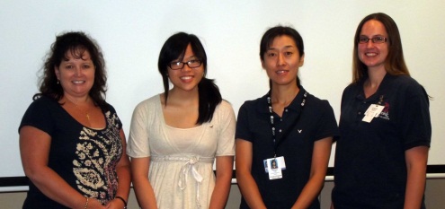
The 2013 Wiegand Intern was Wei Mon Lu a graduate of Serra Catholic High School. Wei is attending Carnegie Mellon University in the Fall. Her internship was in the Plastic Surgery Lab under the mentorship of Dr. Kacey Marra. Pictured are (L to R): Dr. Marra, Wei Mon Lu, Wakako Tsuji, and Jolene Valentin.
University of Pittsburgh Cancer Institute International Academy Scholars Present Projects
 Fifty-six students from around the world participated as scholars in this summer’s University of Pittsburgh Cancer Institute (UPCI) International Academy, a program designed to let students work directly with researchers in basic, translational, and clinical research experiential programs to develop, nurture, and support their interest and aptitude in pursuing careers in biomedicine and cancer. On the final day of the program, students presented findings about the work they did this summer.
McGowan Institute for Regenerative Medicine affiliated faculty member Michael Lotze, MD, UPCI International Academy Program director, as well as other UPCI faculty, staff, and academy scholars participated with the presenting students.
The UPCI International Academy is designed to give students real-world experience in working alongside scientists and cancer clinicians. The goal of the program is to teach students the scientific hallmarks of cancer, helping them learn how to speak the language of science, and promoting science as a performing art.
The program began 5 years ago with 5 participants and has grown this summer to 56 scholars. For the first time in the program, international students also were admitted, including several from Kazakhstan and Germany. Although half hale from the greater Pittsburgh area, many of them are from groups previously under-represented in the biomedical sciences as well as scholars from Hawaii, Minnesota, Vermont, New Jersey, Texas, Virginia, and Maryland.
The students spent the summer working in one of five locations. These included:
- Hillman Cancer Center (cancer biology)
- Women’s Cancer Research Center at Magee-Womens Research Institute (women’s cancers)
- Starzl Biomedical Science Tower (tumor immunology)
- Biomedical Science Tower III (drug discovery, systems, and computational biology)
- Offices at Baum (computer sciences, biological sciences, and biomedical informatics).
Illustration: University of Pittsburgh Cancer Institute.
AWARDS AND RECOGNITION
Jack Hughston Award Received by Dr. Freddie Fu
 McGowan Institute for Regenerative Medicine faculty member Freddie Fu, MD, and Carola Van Eck, MD, PhD, an orthopaedic resident from The Netherlands who came to UPMC and the University of Pittsburgh to learn from Dr. Fu and other sports-medicine experts, are part of a research team presented in Chicago with the prestigious Jack Hughston Award for the outstanding sports-medicine paper of 2012 published in the American Journal of Sports Medicine.
In the paper, Drs. Fu and Van Eck and three researchers from Slovenia – principal investigator Mohsen Hussein, MD, Andrej Cretnik, MD, PhD, and Dejan Dinevski, PhD – concluded that anatomic reconstruction techniques of the knee’s anterior cruciate ligament (ACL) were superior to reconstructing the ACL using non-anatomical drilling techniques. Dr. Fu traveled to the Artros Center for Orthopaedic Surgery and Sports Medicine in Ljubljana, Slovenia, to collaborate with Dr. Hussein, formerly a UPMC Sports Medicine research fellow.
It is the first time a Pitt or UPMC representative has received the decade-old Hughston Award, named for the surgeon who founded the American Journal of Sports Medicine. It was presented during the 2013 convention of the American Orthopaedic Society for Sports Medicine (AOSSM) at the Sheraton Chicago Hotel and Towers.
“It is definitely a tremendous honor for our program and department to win this prestigious award, named for a leader in sports medicine and a surgeon I greatly admired. In addition to expertise in the examination room, hospital room, and operating room, we place great importance on research and education in our practices in sports medicine and the Department of Orthopaedic Surgery. As a result, we have the opportunity to train and collaborate with great people around the world,” said Dr. Fu, chair and David Silver professor of the Pitt Department of Orthopaedic Surgery, and the head team physician for Pitt athletics since 1986. “We have worked tirelessly and passionately to pioneer anatomical ACL reconstruction techniques and make ACL surgery better for the patient. We believe this study shows that anatomical reconstruction will result in the best outcomes for our patients.”
 Regenerative Medicine Podcast Update
The Regenerative Medicine Podcasts remain a popular web destination. Informative and entertaining, these are the most recent interviews:
#126 –– Dr. Jeffrey H. Stern is the Director of Translational Research at the
Neural Stem Cell Institute (NSCI) of New York. NSCI is dedicated to developing regenerative stem cell therapies for various diseases of the central nervous system Dr. Stern discusses his retinal and brain stem cell research.
Visit www.regenerativemedicinetoday.com to keep abreast of the new interviews.