What’s Happening At The McGowan Institute?
August 2012 | VOL. 11, NO. 8 | www.mcgowan.pitt.edu
Regenerative Medicine Projects: 3-D Tissue Chips
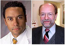
 McGowan Institute for Regenerative Medicine associate director Rocky Tuan, PhD, and fellow researchers at the University of Pittsburgh School of Medicine have been awarded federal grants to create micro-models of an arthritic joint and the liver as part of a national effort to build 3-D chips of cells and tissues that could provide a more rapid and accurate method of predicting toxicity of experimental therapies, as well as foster greater understanding of myriad diseases.
Of the 17 projects being funded by the National Institutes of Health (NIH), two will be led by Pitt researchers and could receive more than $10 million over the next 5 years. NIH plans to commit up to $70 million over 5 years for the Tissue Chips for Drug Screening program, which was launched by its new National Center for Advancing Translational Sciences (NCATS). Other awardees include Johns Hopkins University, Harvard University, and Duke University.
“Tissue chips could provide a more accurate and less expensive way of testing new drugs and reduce our reliance on animal studies, which often don’t reliably reflect toxicity profiles later seen during human testing,” noted Arthur S. Levine, M.D., senior vice chancellor for the health sciences and dean, Pitt School of Medicine. “It is terrific that our stellar scientists will be able to build two of these chips here and contribute to the evolution of drug testing.”
The Pitt projects are:
- 3-D Micro-Arthritic Joint System: Dr. Tuan, the Arthur J. Rooney Sr. Professor of Sports Medicine and executive vice chair for research, Department of Orthopaedic Surgery, will lead a team to create a tissue chip that includes stem cell-produced bone and cartilage cells that simulate joint surfaces to better understand how arthritis develops and how to prevent it.
- “This system will allow us to explore the effects of not only inflammatory molecules and the wear-and-tear of aging on the entire joint, but also mechanical injuries, such as a hit or a sprain, both immediately and over time in molecular detail, which is not feasible with existing techniques,” said Dr. Tuan, who also is director of the Center for Military Medicine Research and director of the Center for Cellular and Molecular Engineering in the Department of Orthopaedic Surgery.
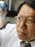
- 3-D Micro-Liver: D. Lansing Taylor, PhD, Allegheny Foundation Professor of Computational and Systems Biology, and director, University of Pittsburgh Drug Discovery Institute, will lead a team at Pitt and Massachusetts General Hospital to create a three-dimensional microfluidic structure made entirely of human cells that will mimic the acinus, the smallest functional unit of the liver. The team also will develop a panel of sentinel “biosensor cells” that will indicate liver toxicity with exposure to different drugs.
“The current gold standard of testing is not very gold,” Dr. Taylor said. “In humans, the liver plays a key role in processing drugs, and many experimental agents have failed in the late stages of human testing because preclinical studies didn’t predict their impact on the liver, along with other organs. This project aims to solve that problem so that we have greater success in drug discovery and development.”
According to NIH, the Tissue Chips program is the result of collaboration between NIH, the Defense Advanced Research Projects Agency (DARPA), and the U.S. Food and Drug Administration, and was established by the NIH’s Common Fund and the National Institute of Neurological Disorders and Stroke.
“Serious adverse effects and toxicity are major obstacles in the drug development process,” said Thomas R. Insel, M.D., NCATS acting director. “With innovative tools and methodologies, such as those developed by the Tissue Chips program, we may be able to accelerate the process by which we identify compounds likely to be safe in humans, saving time and money, and ultimately increasing the quality and number of therapies available for patients.”
Drs. Tuan and Taylor’s projects also will receive support from the University of Pittsburgh Clinical and Translational Science Institute.
SCIENTIFIC ADVANCES
Less Invasive Spinal Surgery Helps Patients Recover Quicker
 At the UPMC Department of Neurosurgery, spine specialists employ minimally invasive techniques and state-of-the-art technologies to treat a range of spine conditions. McGowan Institute for Regenerative Medicine affiliated faculty member David Okonkwo, MD, PhD, assistant professor with the Department of Neurological Surgery/University of Pittsburgh Medical Center, director of Neurotrauma and of the Spinal Deformity Program, clinical director of the Brain Trauma Research Center, and most recently, the associate director of the Center for Injury Research and Control, is teamed with Adam Kanter, MD, the neurosurgeon at UPMC pioneering the minimally invasive technique in Pittsburgh.
Normal spinal surgery involves making multiple incisions, and the operation moves through a larger muscular area. Bone areas have to be removed, and there can be more than a liter of blood loss, said Dr. Kanter to Sanjena Sathian of the Pittsburgh Post-Gazette. With the new technique, which approaches the spine with a “lateral” incision from the patient’s side below the ribs, patients lose about 2 percent of that amount. Patients do not have rods placed along their spine, and they don’t wear a brace afterward.
Dr. Okonkwo said the new approach changes the way he can treat patients over the age of 70 who have scoliosis. For older patients, “it’s a completely different sport,” he said, explaining that elderly patients may already have medical problems and can’t always risk going through a complicated medical procedure.
“There’s no substitute for youth. A 20-year-old who gets a major operation is far more likely to go through without major problems than an 80-year-old,” he said. Minimally invasive spine surgery utilizes advanced techniques and technology to treat a patient’s spinal condition without causing undue injury to the surrounding soft tissues. Computer-assisted technology (such as computer navigation and nerve monitoring) and highly specialized tools and instrumentation provide exceptional visualization and control during these procedures.
Advantages of minimally invasive spinal surgery include:
- Smaller surgical incisions
- Less soft tissue damage
- Reduced blood loss
- Less post-operative pain
- Quicker recovery
- Less scarring
- Improved function
UPMC’s minimally invasive spine surgery specialists are part of a multidisciplinary team that evaluates each patient to help determine the most appropriate intervention. Surgery is recommended only after more conservative medical approaches have failed to provide adequate results for patients seeking pain relief, increased mobility, and improved quality of life.
Regenerative Medicine: Blocking Key Protein Slows Aging
 According to McGowan Institute for Regenerative Medicine affiliated faculty members Paul Robbins, PhD, and Simon Watkins, PhD, and McGowan Institute for Regenerative Medicine faculty members Johnny Huard, PhD, and Donna Stolz, PhD, and other researchers at the Pitt’s School of Medicine, blocking a protein that regulates the activity of certain genes slowed the aging process in a mouse model of premature aging, as well as in healthy mice, The findings, published in the Journal of Clinical Investigation, could lead to drugs that prevent cellular damage due not only to growing old, but also to cancer and diseases caused by abnormal DNA repair activity.
Aging is thought to be the result of accumulated cellular damage, including DNA damage, but the biological mechanisms that drive aging in response to damage are not understood, said senior author Dr. Robbins. His team studied NF-kappa B, a protein involved in turning certain gene activity on and off in response to inflammation, stress, and cellular damage.
“Other studies have shown that NF-kappa B activity is elevated in aging tissues,” Dr. Robbins said. “We examined whether this held true for mice with progeria, a disease of accelerated aging, and what would happen if we blocked NF-kappa B activation.”
The researchers found that a higher percentage of cells contained activated NF-kappa B in old and progeroid mice than in healthy adult mice. Age-related activation of NK-kappa B is stochastic, meaning it happens in some but not all cells, they said.
Altering expression of NF-kappa B slightly or blocking its activation with chemicals led to a delay in the onset and reduction in severity of age-related changes in tissues, including muscle, liver, kidney, and the nervous system. Researchers also found that inhibiting the protein reduced free radical-induced oxidative damage.
“It’s possible that as we age, NF-kappa B becomes activated by accumulation of cellular damage, and that in turn increases the production of free radicals, resulting in more cell damage,” Dr. Robbins said. “An agent that blocks this protein could be used to slow down aging and also to treat certain cancers and diseases such as xeroderma pigmentosum, which are characterized by altered DNA repair activity.”
Abnormal Chromosome Count May Protect Liver Cells from Injury
 The majority of liver cells contain an abnormal number of chromosomes, but these cells still retain their ability to divide. Researchers have long puzzled over why liver cells have unusual chromosome numbers, a state known as aneuploidy, and whether this genetic variation provides some benefit.
To test whether aneuploidy protect liver cells against liver injury, McGowan Institute for Regenerative Medicine faculty member Andrew Duncan, PhD, assistant professor in the University of Pittsburgh’s Department of Pathology, Division of Experimental Pathology, and colleagues at Oregon Health and Science University, Portland, Oregon, examined a mouse model of liver disease caused by deficiency of fumarylacetoacetate hydrolase (Fah). The researchers knew that if these mice acquire a second mutation in proteins that regulate FAH, these mutations provide a selective advantage and protect from liver damage.
To determine whether aneuploidy might contribute to these protective mutations, the Duncan team analyzed chromosomes from the mice. They found a striking prevalence in aneuploidy that lead to protective mutations in all mice that became completely resistant to liver injury.
The team’s results, presented in The Journal of Clinical Investigation, suggest that the selection of a specific aneuploid mutation can result in the adaptation of liver cells to chronic liver injury.
$4.5 Million Grant Obtained to Study Potential Improvements for Wheelchair Users
 McGowan Institute for Regenerative Medicine affiliated faculty member Michael Boninger, MD, professor and chair, Department of Physical Medicine and Rehabilitation, University of Pittsburgh School of Medicine, associate dean of medical student research in the School of Medicine, as well as the director of the University of Pittsburgh Model Center on Spinal Cord Injury, and researchers from Pitt’s School of Medicine and UPMC will lead a 5-year, multi-site project aimed at improving the lives of people with Spinal Cord Injuries (SCI).
The study will use Internet-based training and group sessions to hone the skills of wheelchair use and prevent wheelchair failures. This research will involve more than 500 participants over 4 different sites, making it one of the largest studies of its kind.
Among the other groups involved in the research are:
- the Northern New Jersey Spinal Cord Injury System, a cooperative effort of the Kessler Foundation, the Kessler Institute for Rehabilitation, and the University of Medicine and Dentistry of New Jersey;
- the Midwest Regional Spinal Cord Injury Care System, which brings together Northwestern University’s Feinberg School of Medicine and the Acute Spinal Cord Injury Program at Northwestern Memorial Hospital, plus the Spinal Cord Injury Rehabilitation Program at the Rehabilitation Institute of Chicago; and,
- the University of Miami Miller School of Medicine’s Department of Rehabilitation Medicine and The Miami Project, along with Jackson Memorial Hospital forming the South Florida Spinal Cord Injury System.
“This grant will start to tackle problems related to insurance cutbacks that have negatively impacted individuals with Spinal Cord Injuries,” said Dr. Boninger. “Because they spend less time in the hospital after their injuries, they never learn how to effectively use and maintain their wheelchairs. We need an effective, low-cost way to provide people with training that maximizes their independence – this study tackles that problem.”
Through the efforts of Pitt’s Department of Physical Medicine and Rehabilitation; the School of Health and Rehabilitation Sciences; the UPMC Rehabilitation Institute; and the Human Engineering Research Laboratories – a joint Pitt, UPMC and VA project – Pittsburgh has become nationally and internationally recognized for its expertise in technologies for persons with disabilities.
The latest grant – from the National Institute for Disability and Rehabilitation Research — comes barely 2 months after a published Pitt-UPMC study, with Dr. Boninger as the senior author, found 52 percent of people with SCI required wheelchair repairs in the preceding 6 months. Many of the wheelchair users who needed repairs experienced adverse consequences, the study also found. The same set of Pitt-UPMC researchers also have a 2012 study paper accepted for publication finding a relationship between the ability to perform wheelchair skills – after training – and higher life satisfaction and community participation.
Regenerative Medicine: Nerve Regeneration Technology
 A team led by McGowan Institute for Regenerative Medicine faculty member Kacey Marra, PhD, associate professor of plastic surgery and bioengineering and the Laboratory Director for the Plastic Surgery Research Laboratory at the University of Pittsburgh, is about to launch an intensive, 6-month translational research effort to develop a commercialization strategy for a novel nerve regeneration treatment, thanks to a grant from the National Science Foundation Innovation Corps (I-Corps).
The $50,000 grant will enable the research team from Pitt’s McGowan Institute and Swanson School of Engineering’s Department of Bioengineering to develop a business model for an effective long-gap peripheral nerve repair system with the potential to successfully repair conditions from diabetic neuropathy to battlefield wounds.
The NSF I-Corps program is a highly selective, year-old program and this marks the University’s first I-Corps grant. Because the grant will help to develop the translational and commercialization aspects of the technology, the Pitt team is distinctively structured with an entrepreneurial component to match the NSF goals. Previous funding for this research had been provided by the NSF, the American Association of Hand Surgeons, and the Department of Defense.
Founded in 2012, the Department of Plastic Surgery at Pitt’s School of Medicine builds upon the University’s cutting-edge research and technology, which has advanced clinical areas such as hand and face transplantation, nerve regeneration, and wound healing.
Yen-chih Lin, PhD, serves as Entrepreneurial Lead and has worked in the Marra Laboratory for several years, and has extensive experience in nerve guide fabrication, microsphere fabrication for drug delivery, rat sciatic nerve defect models, non-human primate median nerve defect models, and histological analysis.
The Industrial Mentor is Pratap Khanwilkar, PhD, MBA, Coulter Program Director and professor in the Swanson School of Engineering’s Department of Bioengineering, and Executive-In-Residence with the University’s Office of Technology Management. He has extensive experience in translating research into companies, having obtained seven patents and started six companies, with three operating and generating revenue. One start-up which Dr. Khanwilkar led as founder and CEO for 12 years evolved to a publicly-traded company.
“Nerve repair has improved over the past decade, but one of our challenges remains the regeneration of peripheral nerves over gaps greater than 3 centimeters,” Dr. Marra explains. “Although a seemingly small distance, this represents a chasm that prevents effective treatment of many severed nerve conditions. Our translational research will lead to the development of a commercialization pathway of a biodegradable nerve guide to promote both motor and sensory nerve repair over these long gaps, and for potentially other applications of our proprietary technology.”
“Thanks to the NSF I-Corps grant, we’ll be able to push our decade-long research toward a viable drug-delivery system and thereby address a serious unmet need for clinical care of diseased and damaged nerve tissue.”
The team’s research has advanced from small animal testing and is now being evaluated in larger animals as a precursor to human clinical trials, which Dr. Marra expects within 2 years.
AWARDS AND RECOGNITIONS
McGowan Institute for Regenerative Medicine Affiliated Faculty Member Wins Prestigious Medal
 McGowan Institute for Regenerative Medicine affiliated faculty member Anna Balazs, PhD, distinguished professor of Chemical and Petroleum Engineering at the University of Pittsburgh and also the current Robert Von der Luft professor in that department, has been selected as the 5th recipient of the South Dakota School of Mines and Technology “Mines Medal.” This award recognizes the overall contributions of Dr. Balazs’ career, her influence in engineering or science, and the significance of extraordinary, meritorious, or prestigious contributions toward resolution or understanding of the technological challenges that impact society. Candidates for the Mines Medal:
- show evidence of being transformational individuals, often as rising or confirmed leaders in their fields.
- are scientific or technical leaders who motivate and inspire.
- have specific accomplishments in an appropriate field.
- further the frontiers and expand the boundaries of engineering or science.
The Mines Medal, initiated in 2009, is a national award given annually by the South Dakota School of Mines and Technology to honor engineers and scientists who have demonstrated exceptional leadership and innovation. The award highlights the significant role these individuals play to ensure the United States’ global preeminence in engineering and science.
The Mines Medal Award medallion includes images of the Homestake Gold Mine, a Black Hills grape leaf, and rays of sun emanating from a silhouette of the Black Hills. These traditional symbols of the Black Hills reflect the School of Mines’ longstanding connections to the Homestake Mine in Lead, South Dakota, including the university’s current leadership role in the Sanford Underground Laboratory at Homestake, and the beautiful Black Hills of South Dakota that the School of Mines has called home for 126 years. Materials used in the medallion include 10kt gold and 12kt Black Hills gold in total amount equivalent to one ounce of 24kt gold, and copper and silver.
Illustration: South Dakota School of Mines and Technology (medal image).
Science of the Olympics, Paralympics
 McGowan Institute for Regenerative Medicine affiliated faculty member Rory Cooper, PhD —University of Pittsburgh FISA and Paralyzed Veterans of America Chair and Distinguished Professor in the Department of Rehabilitation Science and Technology, director of the University’s Human Engineering Research Laboratories (HERL), and professor of bioengineering, mechanical engineering, physical medicine and rehabilitation, and orthopaedic medicine at Pitt—was interviewed by NBC regarding biomedical science and biomechanical engineering and technology surrounding athletes in the 2012 Olympic and Paralympic Games.
Dr. Cooper was featured in three online NBC videos as part of the network’s coverage of the Games. The 5-minute educational videos, part of NBC’s “Science of the Olympics” series, highlight athletes including Oscar Pistorius, double-amputee sprinter, and Jenny Simpson, a steeplechaser turned 1,500-meter runner.
“I hope that when people see these programs,” Dr. Cooper said, “it will encourage more people from underrepresented minorities, especially people with disabilities, to pursue careers in science, technology, engineering, and mathematics.”
Dr. Cooper and Pitt doctoral student and HERL physical therapist Justin Laferrier are featured in the following three NBC videos:
- “Science of the Summer Olympics: The Strength and Flexibility of Oscar Pistorius”
- “Science of the Summer Olympics: The Impact of Jenny Simpson”
- “Science of the Summer Olympics: Engineering for Mobility”
“I am really pleased to see the Paralympics get this level of attention,” said Dr. Cooper, a former bronze medalist at the 1988 Seoul Paralympics and recently a five-time gold medalist at the National Veterans Wheelchair Games. “The athletes are truly Olympians and have achieved amazing heights of athleticism.”
The idea for the video series was proposed early this year at a national meeting of the American Association for the Advancement of Science, where Dr. Cooper—there to receive one of two Mentor Award honors—saw a similarly produced educational video on the science behind hockey. Dr. Cooper, with ties to the NSF for roughly 20 years, heard at that meeting about the NSF and NBC partnering on a mirror project involving the Olympics. He suggested the idea of a Paralympics video, and his idea became reality.
Dr. Cooper is a renowned authority on paralyzed veterans, wounded warriors, and on many other military matters who is invited to advise presidential cabinet members and has been honored at a U.S. Marines parade. In addition to being an engineer, a scientist, a researcher, and a professor, he is a veteran and a medal-winning international athlete whom General Mills honored with a photo and write up on a Cheerios cereal box.
 Regenerative Medicine Podcast Update
The Regenerative Medicine Podcasts remain a popular web destination. Informative and entertaining, these are the most recent interviews:
#112 ––Dr. Steven L. Wolf is a research physiologist and professor in the Departments of Medicine and Rehabilitation Medicine at Emory University. Dr. Wolf discusses his work in Regenerative Rehabilitation and the importance of educating clinicians in the field.
Visit www.regenerativemedicinetoday.com to keep abreast of the new interviews.
Publication of the Month
| Authors: | Duncan AW, Hanlon Newell AE, Bi W, Finegold MJ, Olson SB, Beaudet AL, Grompe M. |
| Title: | Aneuploidy as a mechanism for stress-induced liver adaptation. |
| Summary: | Over half of the mature hepatocytes in mice and humans are aneuploid and yet retain full ability to undergo mitosis. This observation has raised the question of whether this unusual somatic genetic variation evolved as an adaptive mechanism in response to hepatic injury. According to this model, hepatotoxic insults select for hepatocytes with specific numerical chromosome abnormalities, rendering them differentially resistant to injury. To test this hypothesis, we utilized a strain of mice heterozygous for a mutation in the homogentisic acid dioxygenase (Hgd) gene located on chromosome 16. Loss of the remaining Hgd allele protects from fumarylacetoacetate hydrolase (Fah) deficiency, a genetic liver disease model. When adult mice heterozygous for Hgd and lacking Fah were exposed to chronic liver damage, injury-resistant nodules consisting of Hgd-null hepatocytes rapidly emerged. To determine whether aneuploidy played a role in this phenomenon, array comparative genomic hybridization (aCGH) and metaphase karyotyping were performed. Strikingly, loss of chromosome 16 was dramatically enriched in all mice that became completely resistant to tyrosinemia-induced hepatic injury. The frequency of chromosome 16-specific aneuploidy was approximately 50%. This result indicates that selection of a specific aneuploid karyotype can result in the adaptation of hepatocytes to chronic liver injury. The extent to which aneuploidy promotes hepatic adaptation in humans remains under investigation. |
| Source: | Journal of Clinical Investigation. 2012 Aug 6. pii: 64026. [Epub ahead of print] |
Grant of the Month
| PI | Rocky S. Tuan | ||
| Title | A Regenerative Medicine Approach for TMJ Meniscus Restoration | ||
| Description |
|
||
| Source | National Institutes of Health – National Center for Advancing Translational Sciences | ||
| Term | 7/24/12 – 6/30/14 | ||
| Amount: | $720,766 total costs for 2 years (includes $224,034 of indirect costs for 2 years) |
Newsletter Comments or Questions: McGowan@pitt.edu