Military personnel are substantially burdened with traumatic bone injury to the extremities, but no ideal therapy is available to regenerate large bone volumes in compromised wounds. These wounds are sub-optimal for regeneration because the vascular damage and immune response provoke oxygen deficiency and inflammation which impair bone growth and drive formation of fibrous tissue.
A Department of Defense (DOD) grant for $2.1M (1.7M direct) over 3 years to accelerate bone healing has been awarded to University of Pittsburgh’s Center for Craniofacial Regeneration (CCR) / McGowan Institute for Regenerative Medicine and the U.S. Army Institute of Surgical Research (ISR). The project’s team of McGowan Institute affiliated faculty members includes:
- Juan Taboas, PhD, Assistant Professor with the Department of Oral Biology in the School of Dental Medicine and the Department of Biomedical Engineering in the Swanson School of Engineering, PI
- Alejandro Almarza, PhD, Associate Professor in Oral Biology in the School of Dental Medicine at the University of Pittsburgh with a secondary appointment in the Department of Bioengineering, CoI
- Yadong Wang, PhD, William Kepler Whiteford Professor in Bioengineering with adjunct positions in Chemical Engineering, Mechanical Engineering and Materials Science, and Surgery at the University of Pittsburgh, CoI
The team’s technology to accelerate bone healing is an off-the-shelf biologic device that can be loaded with minimally manipulated autologous mesenchymal stem cells (MSCs) at the point-of-care. It consists of the following components:
- drugs to control the immune response and promote healing
- hydrogel carrier to deliver the drugs and to control tissue formation by MSCs
- nanoparticles to deliver the drugs over prolonged periods
- porous scaffold to provide mechanical support in large bone defects
These components are assembled in two forms depending on the nature of the bone injury: an injectable hydrogel and an implantable hydrogel infused scaffold. The team expects that an injectable device (components 1-3) will be used by first responders to stabilize bone fragments in comminuted fractures, prevent fibrous tissue ingrowth, and rapidly initiate regeneration. They expect that an implantable device (all components) will be used in the operating theater to promote bone formation and minimize non-union when conventional grafts and bone fillers are contraindicated. They will evaluate bone regeneration over time in two bone injury models in swine, and will test the injectable device in a simulated comminuted fractures of the fibula while the implantable device in large fibular bone defects. Also, they will evaluate bone formation and strength, revascularization and reinnervation, and the local and host immune responses.
Approximately 20% of injured combat personnel suffer extremity bone fracture and loss, of which 80% are open compromised wounds with significant tissue loss post-debridement. The team’s device will minimize the severe morbidity and the treatment cost for wounded military personnel, and will improve their quality of life and return to service. Ultimately, this work will translate to several clinical therapies in the form of pharmacological interventions and therapeutic devices to promote skeletal healing. These will decrease clinical involvedness and impact individual’s lives by speeding skeletal healing, diminishing non-union and tissue fibrosis incidence, and reducing multi-surgery procedures. This work will have a similar strong impact for non-military applications as well.
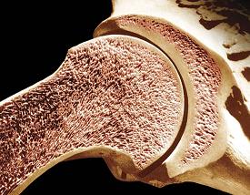
Illustration: Longitudinal section of the humerus (upper arm bone). Dr. Don Fawcett—Visuals Unlimited/Getty Images.
