Although cerebral aneurysms affect a substantial portion of the adult population, the risk of treatment including open brain surgery often outweighs the risks associated with rupture. With increasing numbers of unruptured aneurysms detected using noninvasive imaging techniques, there is an urgent need for a reliable method to distinguish aneurysms vulnerable to impending rupture from those that are presently robust and can be safely monitored. An international research team led by the University of Pittsburgh Swanson School of Engineering recently received a grant from the National Institutes of Health (NIH) to improve risk assessment and treatment of this devastating disease.
Principal investigator of the 5-year, $2,950,622 grant, “Improving cerebral aneurysm risk assessment through understanding wall vulnerability and failure modes,” is McGowan Institute for Regenerative Medicine affiliated faculty member Anne M. Robertson, PhD, the William Kepler Whiteford Professor of Engineering at the Swanson School. McGowan Institute affiliated faculty member Simon Watkins, PhD, Distinguished Professor of Cell Biology and Director of Pitt’s Center for Biological Imaging, is a co-investigator on the study. The R01 grant is funded through the NIH National Institute of Neurological Disorders and Stroke.
“The cells in our blood vessels have a remarkable capacity for rebuilding and maintaining the collagen fibers that give the artery walls their strength. Unfortunately, this natural process can be derailed by the abnormal fluid flow in brain aneurysms, leading to vulnerable walls and rupture,” explained Dr. Robertson. “Understanding the factors that discriminate between robust aneurysm walls with well-organized collagen fibers, and fragile aneurysm walls with diverse changes to the collagen architecture, is essential for improving risk assessment and developing new treatments to prevent rupture.”
To support their work, Dr. Robertson and a multi-disciplinary team of world leaders in aneurysm research from Pitt, Allegheny General Hospital in Pittsburgh, George Mason University in Virginia, University of Illinois at Chicago, and Helsinki University Central Hospital and Kuopio University Hospital in Finland, will analyze brain tissue donated from patients with cerebral aneurysms. Using state-of-the-art facilities for biomechanical analysis and bioimaging, the investigators will specifically look at how and why some patients are naturally able to maintain a healthy aneurysm wall while the walls in other patients weaken, leaving the vulnerable to rupture. They will use computational mechanics to explore the possible multiple mechanisms by which hemodynamics alters the wall and study the mechanisms of structural failure.
“The diverse expertise in our team and our access to an unprecedented number of aneurysm tissue samples enables us to study this disease in an entirely new way,” Dr. Robertson said. “We are also able to leverage computational and experimental tools developed during our prior NIH supported program.”
Other co-investigators on the international team include:
- University of Pittsburgh: Spandan Maiti, PhD, Assistant Professor of Bioengineering
- Allegheny General Hospital: Khaled Aziz, MD, PhD, Department of Neurosurgery
- George Mason University: Juan Cebral, PhD, Professor of Bioengineering, Volgenau School of Engineering
- University of Illinois College of Medicine at Chicago: Fady Charbel, MD, Professor of Neurosurgery and Chief of Neurovascular Section; and Sepi Amin-Hanjani, MD, Professor of Neurosurgery
- Kuopio University Hospital (Finland): Juhana Frösen, MD, PhD, Department of Neurosurgery
- Helsinki University Central Hospital (Finland): Riikka Tulamo, MD, PhD, Department of Vascular Surgery
“Because of the critical importance and delicate nature of the brain, surgical treatment of cerebral aneurysms is avoided unless absolutely necessary. That’s why doctors and surgeons need a more effective way to determine whether a patient with a cerebral aneurysm is at risk for rupture,” Dr. Robertson said. “We expect that by understanding the differences in the vulnerable and robust aneurysm wall, we will be able to improve risk assessment, identify biomarkers of wall fragility, and provide essential knowledge for developing pharmacological treatments to harness and augment the natural repair process of the aneurysm wall.”
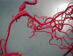
Illustration: Cast of human brain vessels showing cerebral aneurysm in center, taken by Charles Kerber, MD.
Read more…
University of Pittsburgh Swanson School of Engineering News Release
