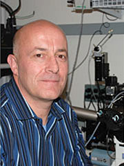
Simon C. Watkins, PhD
Dr. Simon Watkins is the Founder and Director of the Center for Biologic Imaging at the University of Pittsburgh and a member of the Pittsburgh Cancer Institute. He is also a Distinguished Professor and Vice Chairman within the Department of Cell Biology.
Dr. Watkins received his PhD in Neurobiology from Newcastle University in England in 1983. He also holds a BSC in Zoology from Hull University in England. Following his graduate studies, Dr. Watkins was appointed as a Research Fellow at Pasteur Institute in Paris, France, within the Department of Molecular Biology. Immediately prior to arriving at the university, he was a Research Fellow and Research Associate at the Harvard Medical School Dana-Farber Cancer Institute.
The driving forces behind Dr. Watkins’ research are cutting edge optical imaging and their application to studying basic cell biologic processes. The CBI which he founded and directs is a center employing 4 faculty and over 20 staff. The Center builds, tests, and uses cutting edge optical tools for all types of research microscopic imaging in cells, tissues and animals from the single molecule to the whole animal, the goal being to build highly flexible, maximally effective imaging solutions, to be used by academic researchers. In fact a major focus of his career and of the Center is to develop, train and imbue researchers at all levels (undergraduate, student, post-doc and faculty) with a solid understanding (both theoretical and practical) of the power of microscopy.
As a professor of Cell Biology a major focus of his research has been to develop, build, and apply computer aided microscopes and analysis tools for imaging subcellular events at all levels of resolution within fixed and living systems. These include high speed Total Internal Reflection Fluorescence microscopes able to image at 100 frames/second, high speed confocal systems able to collect multicolor 3D stacks in the second timeframe and other prototype confocal systems able to scan very large tissue sections with submicron resolution at very high speed. His achievements in this field led to his promotion to Distinguished Professor in 2014. Most recently he has been developing very high speed deep tissue imaging solutions to collect quantitative images at the diffraction limit of entire tissues including brain. The devices being worked on are effectively 20-30 times faster than a conventional confocal microscope making truly massive scale imaging a possibility. These studies are performed in both living and fixed systems.
Dr. Watkins is the author of over 700 publications and has an H index of 136. View a list of his publications here.
He is an invited reviewer of many journals, including Muscle and Nerve, Journal of Neurological Sciences, Journal of Cell Biology, “Agents and Actions,” American Journal of Pathology, and Journal of Immunology. He is an editor of Current Protocols in Cytometry, Experimental Science and Medicine, and Microscopy Today.

